KEYNOTE SPEAKERS
CV
– Graduated cum laude at University of Chieti, Italy in 1988.
– Former President of the Italian Society of Endodontics.
– Former President of the Congress of the European Society of Endodontology (ESE), 2011.
– Honorary member of the French Society of Endodontics.
– Visiting Professor, International Master in Endodontics and Restorative Dentistry, University of Siena, Italy.
– Founder of the Italian Academy of Microscopic Dentistry.
– Master of Science in Interdisciplinary Dentistry.
– Private clinic devoted primarily to endodontics, pre-prosthetic core build-up, restorative procedures and prosthodontics under operatory microscope, Rome, Italy.
– Author or co-author of numerous research papers on endodontics and post-endodontic core build-up.
– Presenter of numerous lectures and practical workshops in Italy and worldwide on Endodontics, Restorative procedures and use of the operatory microscope in dentistry.
Abstract
Restoration of broken-down teeth remains the final objective of the majority of dental treatments. Root to Crown is a systematic approach to the different scenarios the dentist faces in everyday practice, its objective is to simplify the treatment plan as well as the treatment in vital cases and in non-vital cases. The principle is: saving tooth structure, use simplified adhesive systems and ideal materials. Modern adhesives and composites offer better performance regardless of the fact that vital or non-vital tooth is under treatment.
In the anterior area they offer ideal color integration due to correct translucency and handling properties that, associated with a guided approach for design result is simple yet successful restoration of the esthetic zone. Posteriors are today approached in few simple steps and do not need layering in order do accomplish good combination of form and function. Post-endodontic restoration shows problems and limitations which are very well known. Residual space geometry is not ideal for materials commonly used, it can be not homogeneous and as final result it becomes difficult to adapt prefabricated posts to the residual space. Several evidence state the importance of the coronal restoration of endodontically treated teeth in order to obtain the establishment of the system.
Endodontic treatment seen in its total integration with the restoration tends to be rapid, reliable , and simplified when new materials are used. Importance will be given to the different variables influencing the restoration used interface and the gap formation when posts are used as well as when posts are not used in combination with different materials, adhesives, and endodontic sealers. New composite materials are the ones that play the biggest role in the Post Endo game offering simplicity and predictability. Different modalities and precise approach of post endo restoration will be analyzed from a clinical point of view as well as a microstructural point of view.
CV
– Received DDS degree in 1985 and Specialization in Orthodontics and Dentofacial Orthopedics from Venezuelan Central University, Caracas, Venezuela in 1993.
– Honorary Member Venezuelan Orthodontics Society.
– Instructor professor in Venezuelan Central University (UCV) from 1994 – 1996.
– Collaborating professor in Orthodontics Postgraduate Program, Venezuelan Central University
– International Lecturer in many countries: Mexico – Perú – Brasil – USA – Slovenia – Italy – Vietnam –
Venezuela.
Abstract
Recent research has demonstrated the correlation between occlusal plane and the position and morphology of the mandible. This knowledge has helped us with a better understanding of normal growth, the vertical dimension as a key factor for the normal development of the condyle/ ramus and malocclusions, their correction approach and future stability.
The lecture will focus on interceptive and corrective therapies for the most common malocclusions, such as Class II, Open Bites, Class III, skeletal deformities of facial convexity or concavity, and lateral deviations of the mandible. This approach is based on 3D management of the occlusal plane with emphasis in the vertical dimension with the use of BioMeaw mechanics, which is a combination of Bioprogressive and GEAW principles.

CV
– World-renowned researcher and clinician in Minimal Interventional Dentistry and Cavity Pathology.
– Professor at the University of Queensland, Australia from 2009.
– Former Dean of the Faculty of Dentistry, University of Kuwait in 2012 and University of Sharjah, UAE in 2016.
Visiting Professor of:
+ King College, London.
+ University of Queensland, Australia.
+ University of Malaya, Kuala Lumpur, Malaysia.
+ University of Singapore, Singapore.
+ University of Thammasat, Bangkok, Thailand.
– Dean of the University of Western Australia Dental School.
– Director of the Oral Health Centre Western Australia.
– Member of the State Oral Health Council of Western Australia.
Abstract
Dental caries continues to be a significant public health issue, adversely affecting individual well-being through financial strain, persistent pain, functional difficulties, and decreased quality of life. The emergence of Minimal Intervention (MI) dentistry has transformed caries management, supported by important policy statements from the FDI in 2002, 2012, and 2013.
MI focuses on a patient-centred and non-invasive approach to caries treatment, integrating minimally invasive surgical methods to preserve tooth structure and vitality. This presentation will delve into the clinical applications of MI in caries management, highlighting areas that require additional investigation. Topics of discussion will encompass evaluating patient risk factors, developing customized management plans, addressing both caries and erosion, effectively employing preventive products, and suggesting care strategies for both in-office and at-home settings. Attendees will acquire valuable knowledge about cutting-edge dental materials that can enhance their capacity to provide effective and consistent patient care.
Learning Outcomes:
– Strategies for managing early non-cavitated lesions.
– The synergistic relationship between fluoride and strontium.
– The mechanism of action of CPP-ACP.
– Minimally Invasive restorative techniques.
Topic 2. Know-how on implant surgery & prosthdontics for immediate loading protocol

CV
– Received DDS degree from Kyung Hee University College of Dentistry, Seoul, South Korea in 1984.
– Received PhD degree from Kyung Hee University College of Dentistry, Seoul, South Korea in 1992.
– Received Prof. title from Kyung Hee University College of Dentistry, Seoul, South Korea in 1993.
– Tenure Professor, Department of Prosthodontics, Kyung Hee University College of Dentistry, Seoul, South Korea from 1993-2023.
– Prof. Emeritus, Department of Prosthodontics, Kyung Hee University College of Dentistry, Seoul, South Korea from 2023.
Abstract
Abstract 1: Primary implant stability is a prerequisite for successful osseointegration, and that implant instability results in fibrous encapsulation. A high value of insertion torque (30~50 Ncm) seems to be one of the prerequisites for a successful immediate/early loading procedure.
Techniques for measuring implant stability and osseointegration, including the clinical measurement of cutting resistance during implant placement and removal torque following osseointegration, are discussed.
Two measurement methods at the micromotion-level of an implant are widely used: the self-resonance frequency analysis (RFA) method and the percussion method. The RFA method requires connecting to the implant every time a transducer called SmartPeg is measured, but there is a hassle of specifically selecting and measuring the right one depending on the type of implant. Unlike this RFA method, the percussion method has the advantage of being able to freely measure stability whenever necessary even during the healing period without removing the healing abutment (HA) or upper implant prosthesis. There is already well-known and released AnyCheck (Neobiotech Co. Ltd., Seoul, Korea).
Both measuring instruments are judged to be stable when recording a value of 70 or more as a micromotion value (ISTv).Objectives:
– Understand the requirements on implant surgery for immediate/early loading.
– Understand what are the hazards to the bones that lead to implant failure during implant surgery.
– Ensure that CMI fixation concept is understood and realized.
– Understand and practice Top-Down implant treatment concept.
– Prepare to determine the timing of loading to follow the type of implant rehabilitation.
Abstract 2: Embark on a transformative exploration of stability and aesthetics in full-arch implantology based on Top-Down & Restoration-Driven Treatment Planning. Transitioning to the realm of All-on-4 and All-on-X, where cross-arch splinting emerges as a potential catalyst in minimizing required implants for full-arch cases, I uncover the innovative possibilities on these reductions unleash—reshaping the landscape of implant dentistry. Drawing on extensive experience and research, I share long-term outcomes and successful methodology based on my scientific life over 39 years, offering a comprehensive perspective on achieving stability and aesthetic excellence in full-arch implantology.
CV
– Received DDS degree from University of California, San Francisco, USA in 1993.
– Received MSc degree from School of Dentistry, Loma Linda University, USA in 2003.
– Received Prof. title from Loma Linda University, USA in 2013
– CEO, Wilshire Periodontics and Dental Implant Center.
– Lecturer, School of Dentistry, University of California, Los Angeles, USA.
Abstract
This course will guide attendees on the concept of guided bone regeneration, its history, and the principles that enable it to be a predictable bone augmentation technique. Biomaterials used in the GBR procedure will also be reviewed. Tent Screw techniques on GBR will be reviewed with various clinical cases.
Objectives:
– Learn the history and concept of GBR.
– Learn bone biology is required to understand the GBR principles.
– How to grow bone by using tent screws predictably.
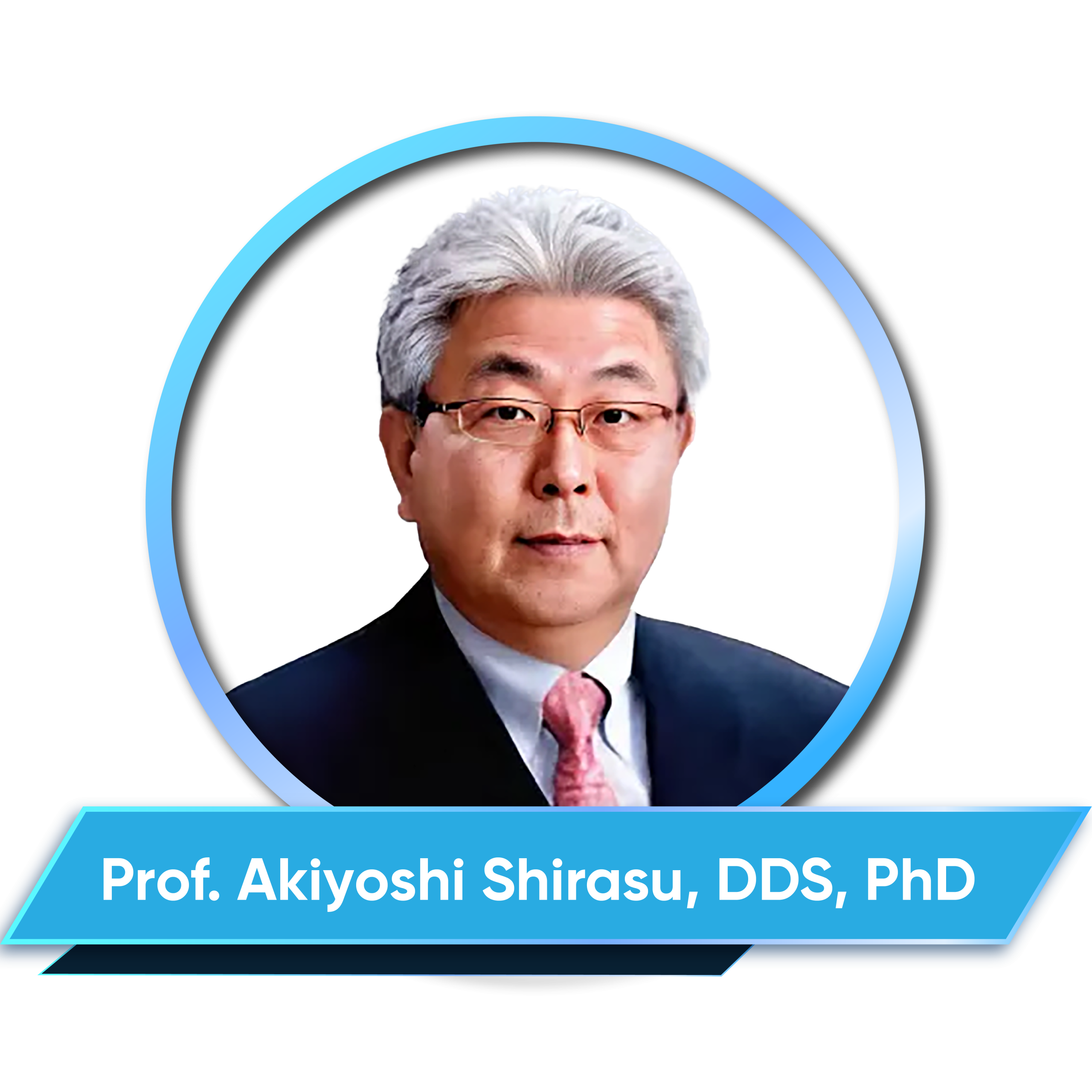

CV
– Received DDS degree from Dental College of Gifu (Now University of Asahi) / Gifu, Japan in 1978.
– Received MSc degree from University of Okayama, Japan in 1987.
– Visiting teaching faculty at Department of Orthodontics, Kanagawa Dental College / Kanagawa, Japan from 2000-2003.
– Visiting teaching faculty at Department of Craniofacial Growth & Development Dentistry / Kanagawa Dental College/ Kanagawa, Japan from 2003-2015.
– Private Dental Clinic in Okayama, Japan from 1981-2009.
– Private Dental Clinic and Office (SHIRASU Dental Office) in Okayama/ Okayama, Japan since 2009.
Abstract
To establish a physiologic and functional occlusion, we must consider the principles of adaptation and compensation, make a precise diagnosis (strategy) based on the concept of malocclusion and a well-defined treatment plan (tactics), and execute the tactics for achieving proper mandibular position, occlusal vertical dimension, occlusal plane inclination, occlusal guidance of each tooth, and stress management.
Orthodontic appliances will not produce effective results if they are used solely as gears or gadgets to move teeth. Achieving normal occlusion for individuals requires mechanics with a treatment system based on proper diagnosis and treatment planning. This time, I would like to introduce my technique for treating malocclusion with TMJ dysfunction utilizing the GEAW system.
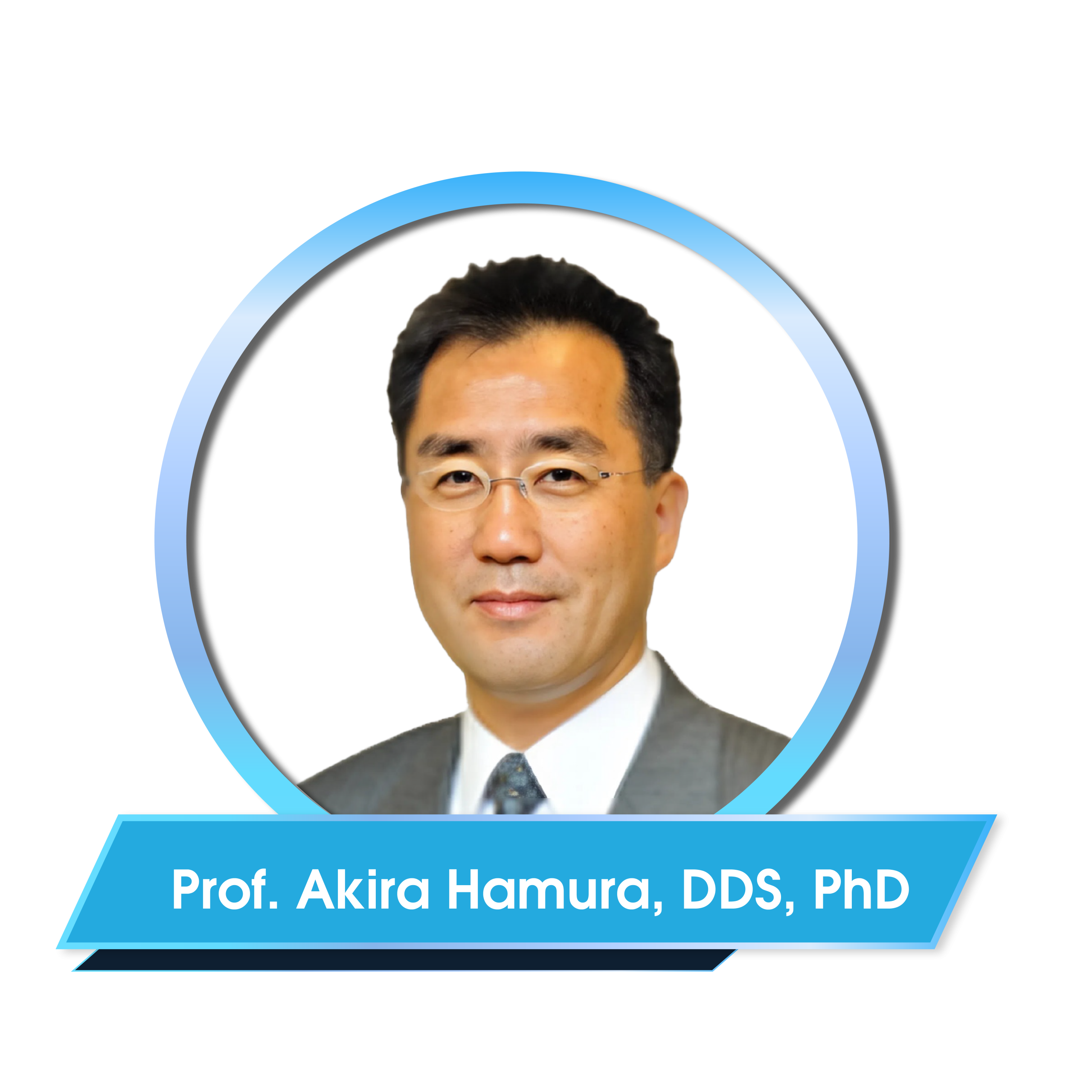
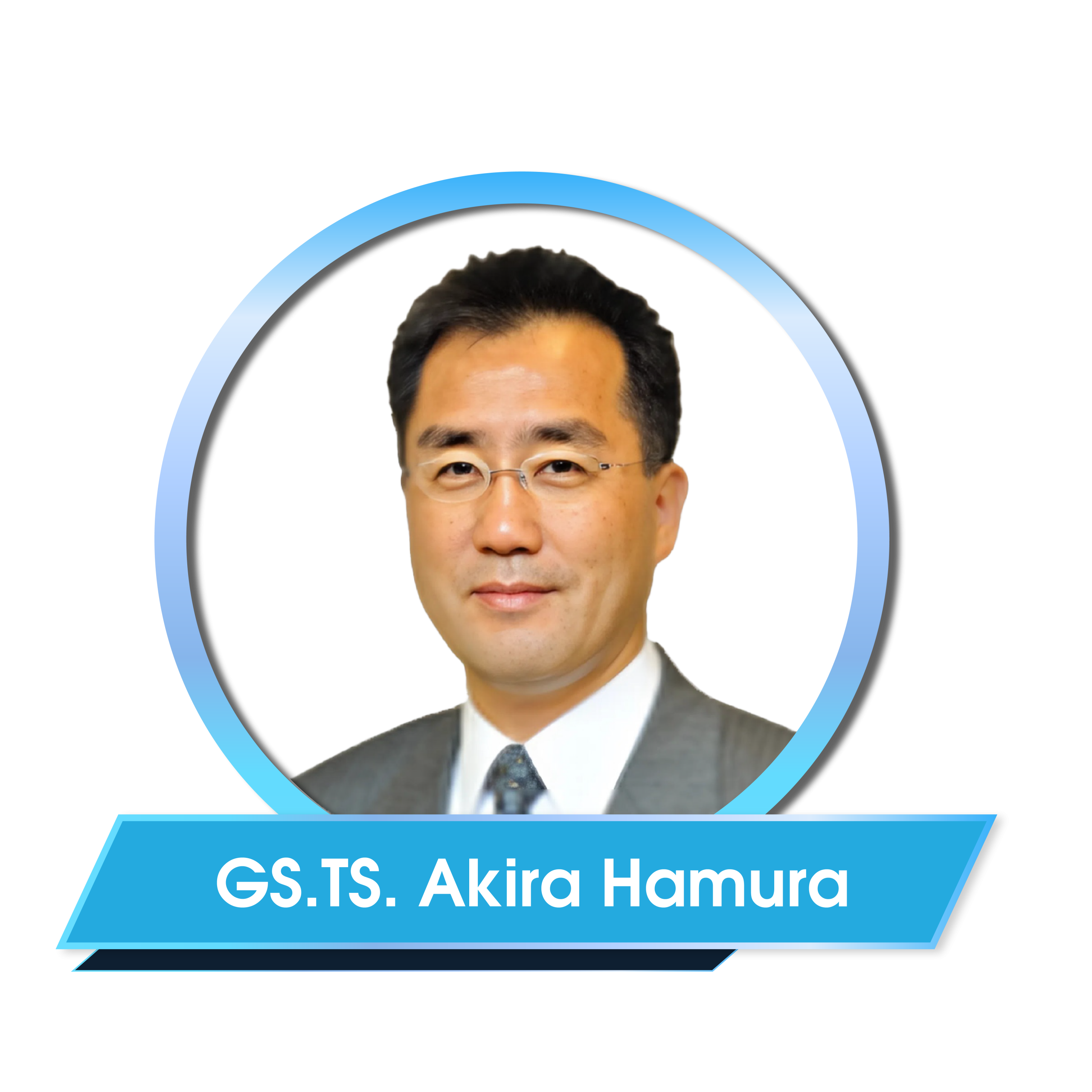
CV
– Received DDS degree from The Nippon Dental University, Japan in 1979.
– Received PhD degree in Prosthodontic from The Nippon Dental University, Japan in 1983.
– Dean and Professor, The Nippon Dental University School of Life Dentistry, Department of Geriatric Dentistry.
– Visiting Professor, Department of Cariology,School of Dentistry, University of Turku, Finland.
– Executive Director of Japanese Society of Geriatric Dentistry.
– Councillor of Japanese Society of Special needs Dentistry.
– Vice Chief Director of Council of Japan Dental Science and Societies.
– Chief Director of Japan – Finland Society for Caries Prevention.
Abstract
In the late of 19th century, it was discovered that Xylitol exists in nature. German Professor Emil Fischer, a Nobel Prize winner in chemistry, was the first to extract xylitol from beech trees. Later, in the 1960s, xylitol began to be produced industrially in Finland, and it became clear that xylitol has a positive effect on oral health. Turku Sugar Studies published in 1975 revealed that chewing xylitol gum after meals was as effective in preventing of caries as replacing all sugar in the diet with xylitol chewing xylitol gum after meals.
In 1986, a clinical trial of xylitol chewing gum was conducted in Ylivieska, Finland, on 11-12 year old children, and xylitol intake reduced the incidence of caries by 30-80%. Furthermore, in 1996, it was revealed that teeth that erupted during or after intake of xylitol had a long-term effect of preventing the onset of caries. In 2000, a field study of schoolchildren in Estonia reported that there was no difference in the effect of xylitol on caries prevention whether it was ingested as chewing gum or candy. In 2013, it was reported that mothers’ use of xylitol chewing gum prevent the caries occurrence in their children. Many studies have been done using xylitol, and several systematic reviews have been reported. From these studies and reviews that habitual intake of approximately 5 g of xylitol per day significantly reduces the incidence of dental caries.
In the presentation, I would like to introduce xylitol research conducted on children and discuss the caries-inhibiting effect of xylitol intake.
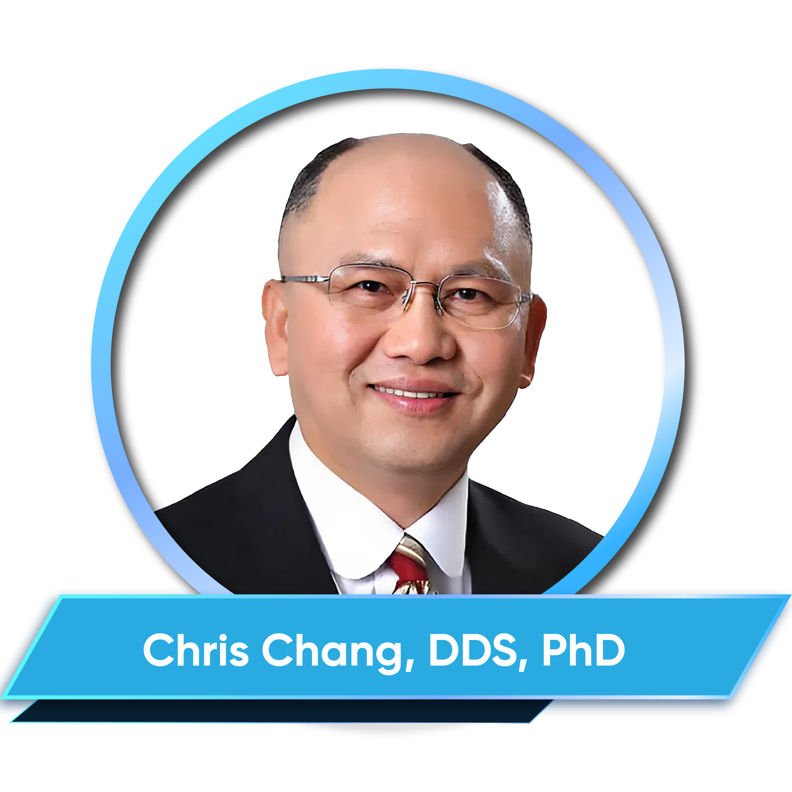
Topic 2: Advanced orthodontics - biomechanics, force control and new approach to complex cases
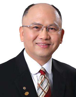
CV
– Received PhD degree in Bone Physiology and Certificate in Orthodontics from Indiana University, USA.
– Founder of Beethoven Orthodontic Center and Newton’s A Inc. in Hsinchu, Taiwan.
– Diplomate of the American Board of Orthodontics.
– Active member of Angle Society-Midwest, USA.
Abstract
With over 23 years of experience in designing and refining miniscrew applications in orthodontics, this presentation introduces six innovative techniques for using miniscrews in four key sites: the infrazygomatic crest (IZC), buccal shelf, incisor region, and ramus. Miniscrews can be placed in one or multiple sites to address sagittal, vertical, and transverse discrepancies, as well as complex impactions. These key applications enhance precision, flexibility, and treatment efficiency, offering reliable solutions for a wide range of challenging orthodontic cases.
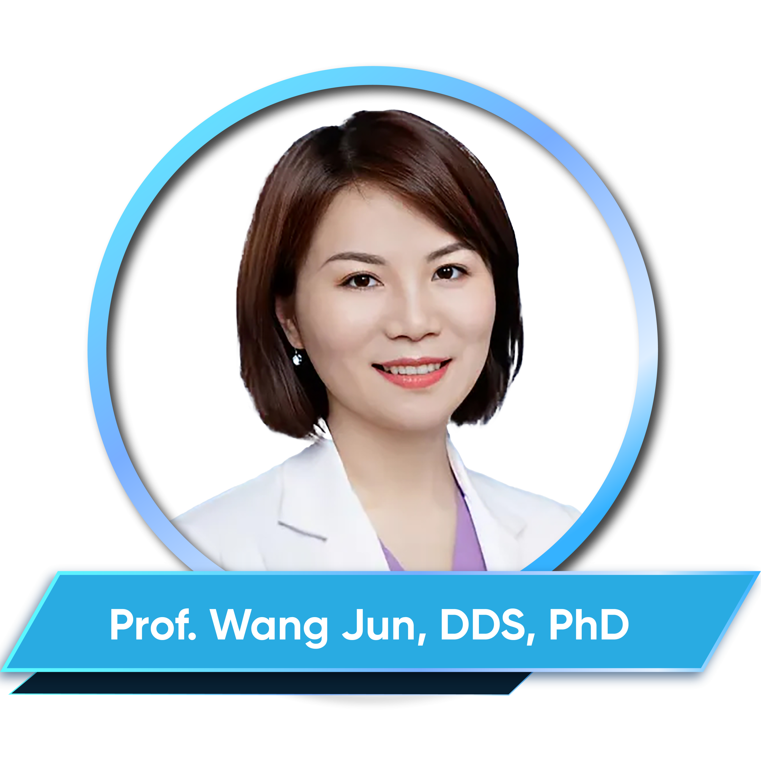
CV
– Director, Professor, Department of Pediatric Dentistry, Ninth People’s Hospital Affiliated to Shanghai Jiao Tong University School of Medicine, China.
– Fellow of the International College of Dentists (ICD), China Section.
– Board member of Pediatric Dentistry Association of Asia.
– Chairman of the Chinese Society of Pediatric Dentistry.
– Clinical Expertise:Specializes in pediatric dental caries.
Abstract
Traditionally, vital pulp therapy (VPT) has been predominantly indicated for endodontic management of immature permanent teeth. However, in recent years, there has been a growing trend toward applying VPT to fully developed permanent teeth. Historically, VPT was only performed on teeth with normal pulps or reversible pulpitis. Nevertheless, accumulating evidence now demonstrates the feasibility of successful VPT outcomes in teeth with irreversible pulpitis or periapical periodontitis. This lecture aims to elaborate on key questions, including which teeth with irreversible pulpitis or periapical periodontitis are suitable for attempted VPT, how to conduct clinical evaluation and perform the procedure, and will systematically address these aspects.
Learning objectives
1. Identify diagnostic criteria and clinical signs that support the use of VPT in cases of irreversible pulpitis and apical periodontitis
2. Recognize key factors influencing VPT success, including vitality assessment, hemostasis, aseptic technique, and material selection.
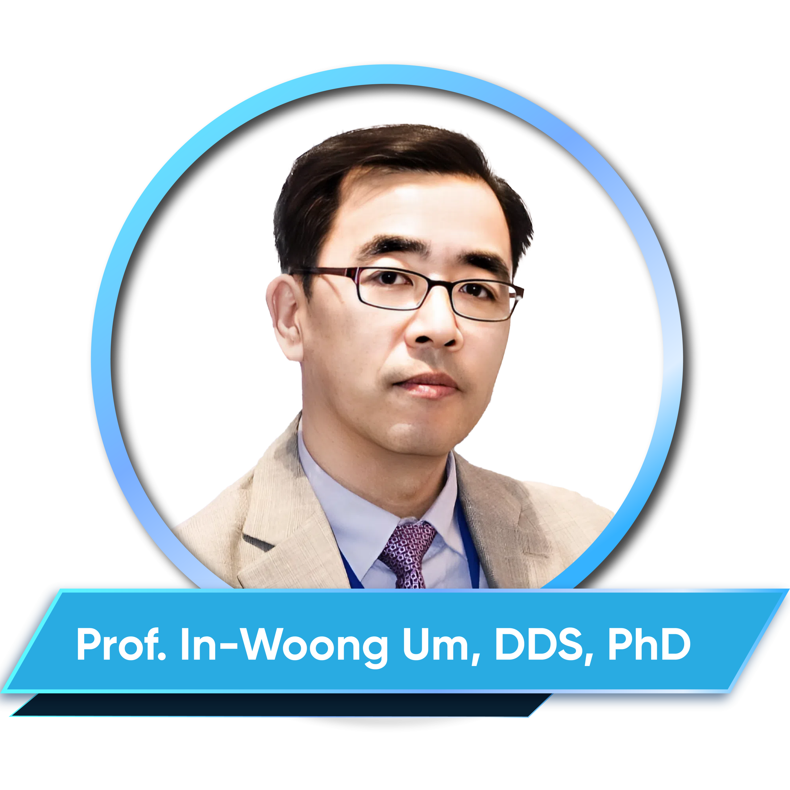
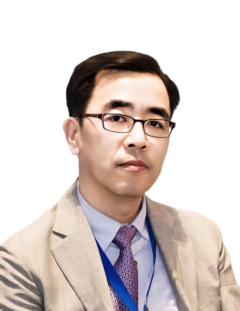
CV
– Received DDS degree from Seoul National University Dental College, Korea in 1984.
– Received PhD degree from Seoul National University Dental College, Korea in 1992.
– Received Prof. title from Chungnam National University College of Medicine, Korea in 1990
– Vice-President of The Korea Academy of Implant Dentistry from 2013-2015.
– Director, The Korean Academy of Implant Dentistry from 2017-2019.
– Former Chairman, Implant Clinical Research Committee, KAOMS
– Director of R&D Institute, Korea Tooth Bank.
– Dentist at Sangryu Dental Clinic, Korea.
Abstract
As the autogenous bone graft still remains “Gold Standard” in implant dentistry, several bone graft substitutes have been developed to come up with “God Standard” in terms of components and function such as osteoinduction, osteoconduction, and osteogenesis.
Based on highly ranked evidence-based literature and qualified certification, I will introduce the bone graft technique using Hu-BT on socket preservation (ridge preservation), ridge augmentation, guided bone regeneration, ridge split, and sinus augmentation as Simplest Bone Graft Technique. Since the introduction of tooth-derived bone graft substitute in 2015 (Hu-BT, Korea Tooth Bank, Seoul, Korea), clinical safety and efficacy has been approved by KFDA and KAHW as similar to the “Gold Standard” in component and function.
In addition, I will provide clinical studies and long-term results on whether we achieve the final goal of bone graft in dental implant as the “Evidence-Proof” of “Successful Implant”.
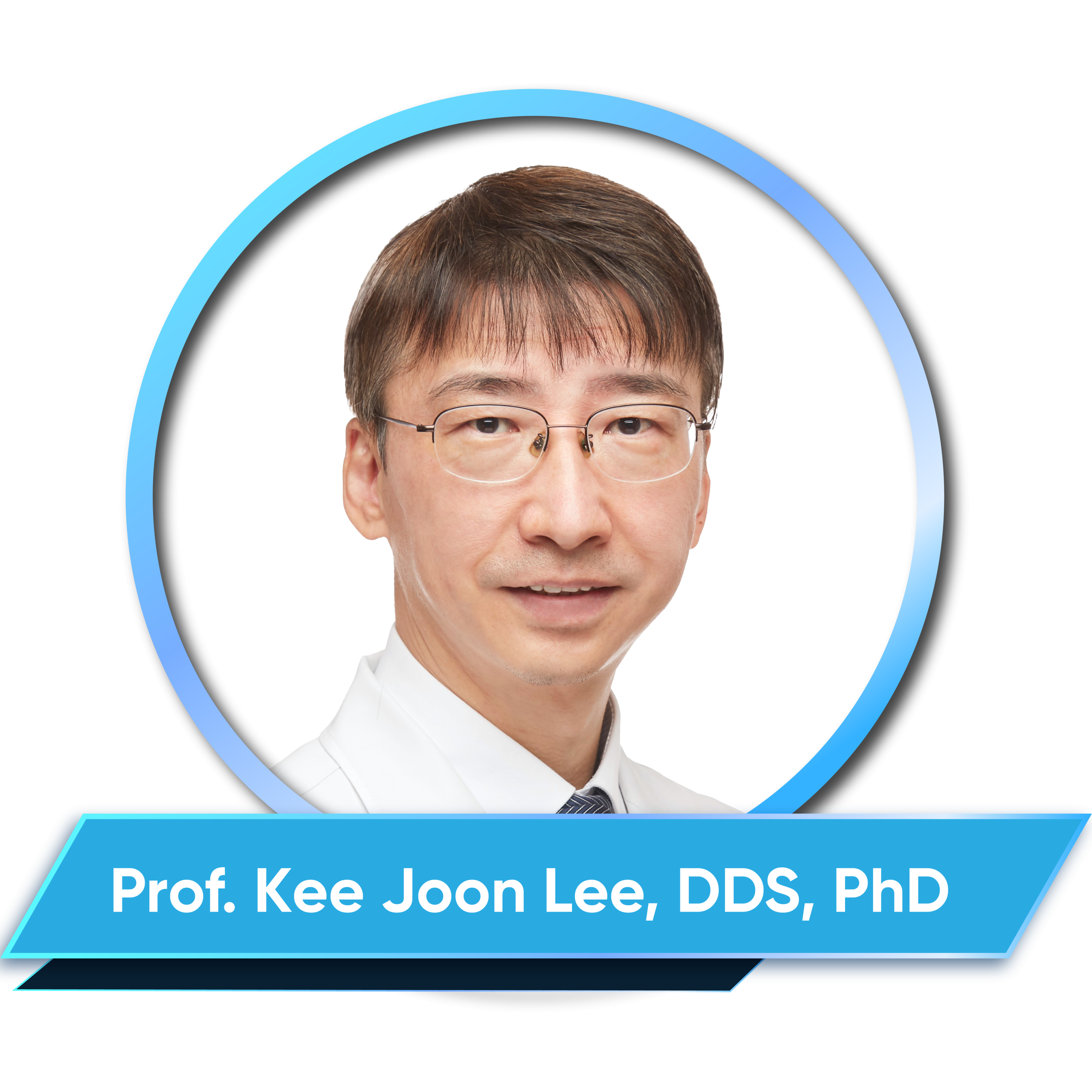
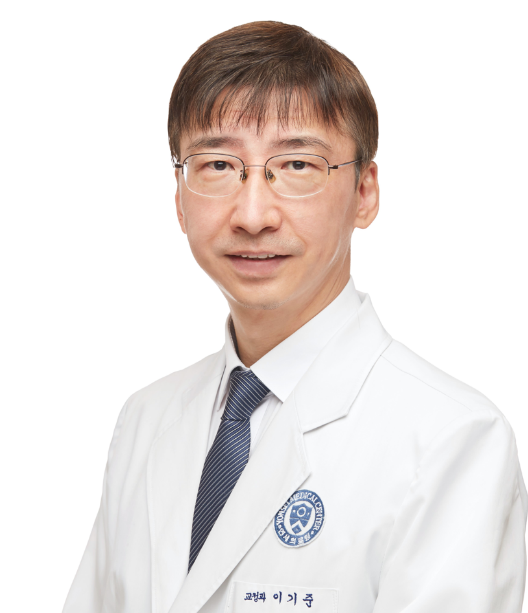
CV
– Received DDS degree from Yonsei University, Seoul, South Korea in 1994.
– Received PhD degree from Yonsei University, Seoul, South Korea in 2004.
– Received Prof. title from Yonsei University, Seoul, South Korea in 2004.
– Visiting scholar, University of Pennsylvania from 2002 to 2004.
– Adjunct professor, University of Pennsylvania, Temple University, USA from 2010 to 2011.
– Visiting scholar, the Children’s Hospital of Philadelphia from 2010 to 2011.
– Professor, Department of Orthodontics, Yonsei University College of Dentistry.
– Former Dean, Yonsei University College of Dentistry, Seoul, South Korea.
Abstract
Conventional orthodontic treatment is conducted by combined use of bracket and wire. Apart from the main advantages of the brackets in three-dimensional control, inherent bracket-wire play may cause unwanted tooth movement within the slot commonly called as round-tripping. Unwanted movement, even in small amount, may be detrimental in serious pathologic situation such as periodontal loss, root resorption and temporomandibular joint disease, as well as significant relapse, where extremely precise tooth movement is indicated. In this scenario, a specific brace that eliminates the possible play is crucial.
In this presentation, recent advances in the bracket-free segmental or total arch movement using force-driven approach in various critical cases will be explained and demonstrated. The essential component includes ribbon arch with various actions in three dimensions, miniscrews and the adequate composite for direct bonding. Latest introduction of the flowable composite with optimal flow would be suitable for this approach with high clinical efficiency. Myofunctional therapy using specific flow is also possible without the need for laboratory procedure. Relevant clinical cases with three-dimensional occlusal aberrations.
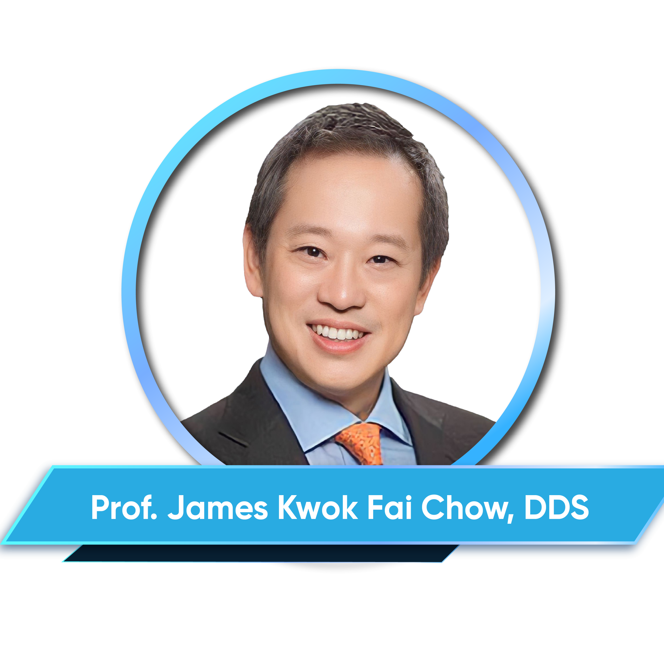
CV
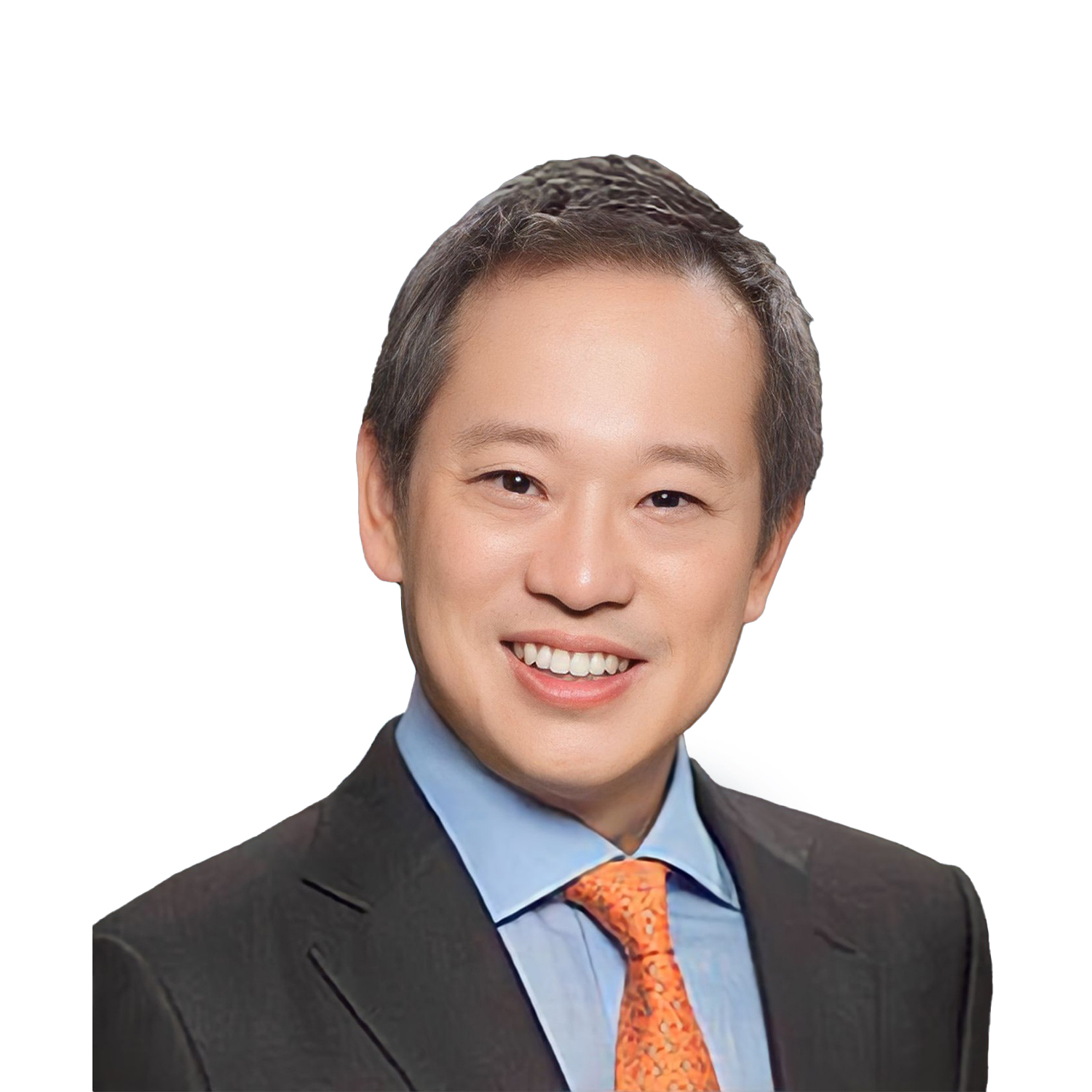
– Received BDS degree from University of Hong Kong in 1988.
– Received MSc degree in Dental Surgery from University of Hong Kong in 1993.
– Received Bachelor of Medicine and Bachelor of Surgery from University of Hong Kong in 1998.
– Fellow of the College of Dental Surgeons of Hong Kong and Hong Kong Academy of Medicine in 2001.
– Diploma in Implant Dentistry, Royal College of Surgeons of England in 2009.
– Fellow of the Royal College of Surgeons of England in 2015.
Abstract
Full-arch implant rehabilitation in patients with severely atrophic maxilla presents significant challenges due to limited bone volume and proximity to critical anatomical structures such as the maxillary sinus, nasal cavity, and orbital floor. Advanced techniques, including zygomatic (bilateral or quad) and pterygoid implants, require exceptional precision to achieve optimal positioning and avoid complications.
Dynamic and robotic computer-assisted implant surgery (CAIS) systems, such as the Dcarer platform, offer real-time image guidance and unparalleled accuracy, enabling clinicians to execute complex procedures with confidence. This one-day course provides a comprehensive exploration of dynamic navigation and robotic-assisted implant placement, focusing on their application in atrophic maxilla cases.
CV
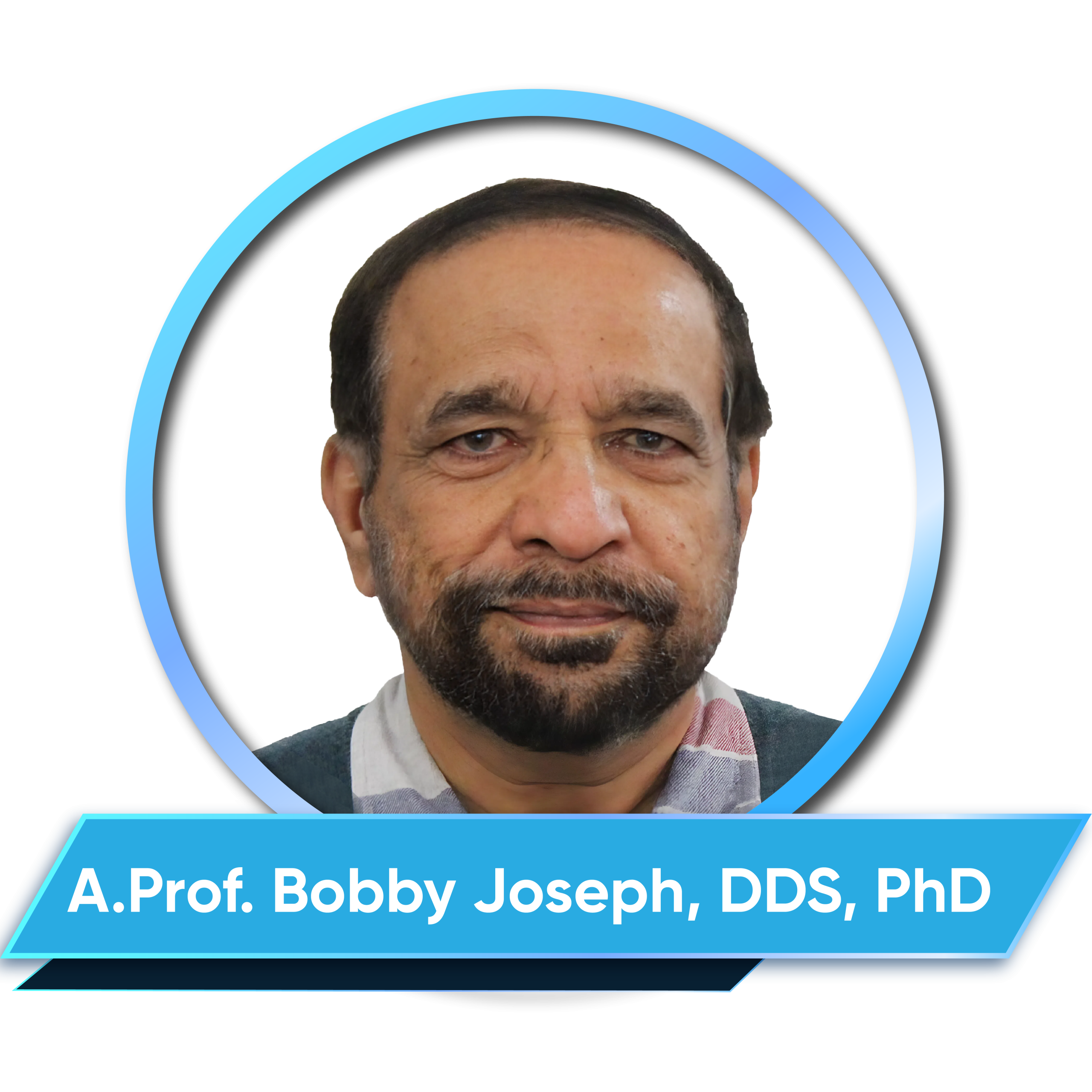
– Received MDS degree from University of Queensland, Australia in 1989.
– Received PhD degree from University of Queensland, Australia in 1994.
– Former Lecturer, University of Queensland, Kuwait University.
– Lecturer, The University of Western Australia, Australia.
Abstract
Despite being a relatively common oral mucosal disease, oral lichen planus (OLP) is still a disease without a clear aetiology or pathogenesis, and with uncertain premalignant potential. While OLP is a chronic inflammatory disorder of unknown aetiology, oral lichenoid reactions (OLR) develop in response to an extrinsic agent.
Accurate diagnosis of OLP is often challenging and emphasis is made on a clinico-pathological correlation in the diagnostic process. OLP pursues a chronic course, in some patients extending over several years. Lesions are generally bilateral, and a wide spectrum of clinical presentations occur. This lecture will examine the current knowledge on the diagnosis and management of OLP and will also focus on the challenges of making an accurate diagnosis.

CV
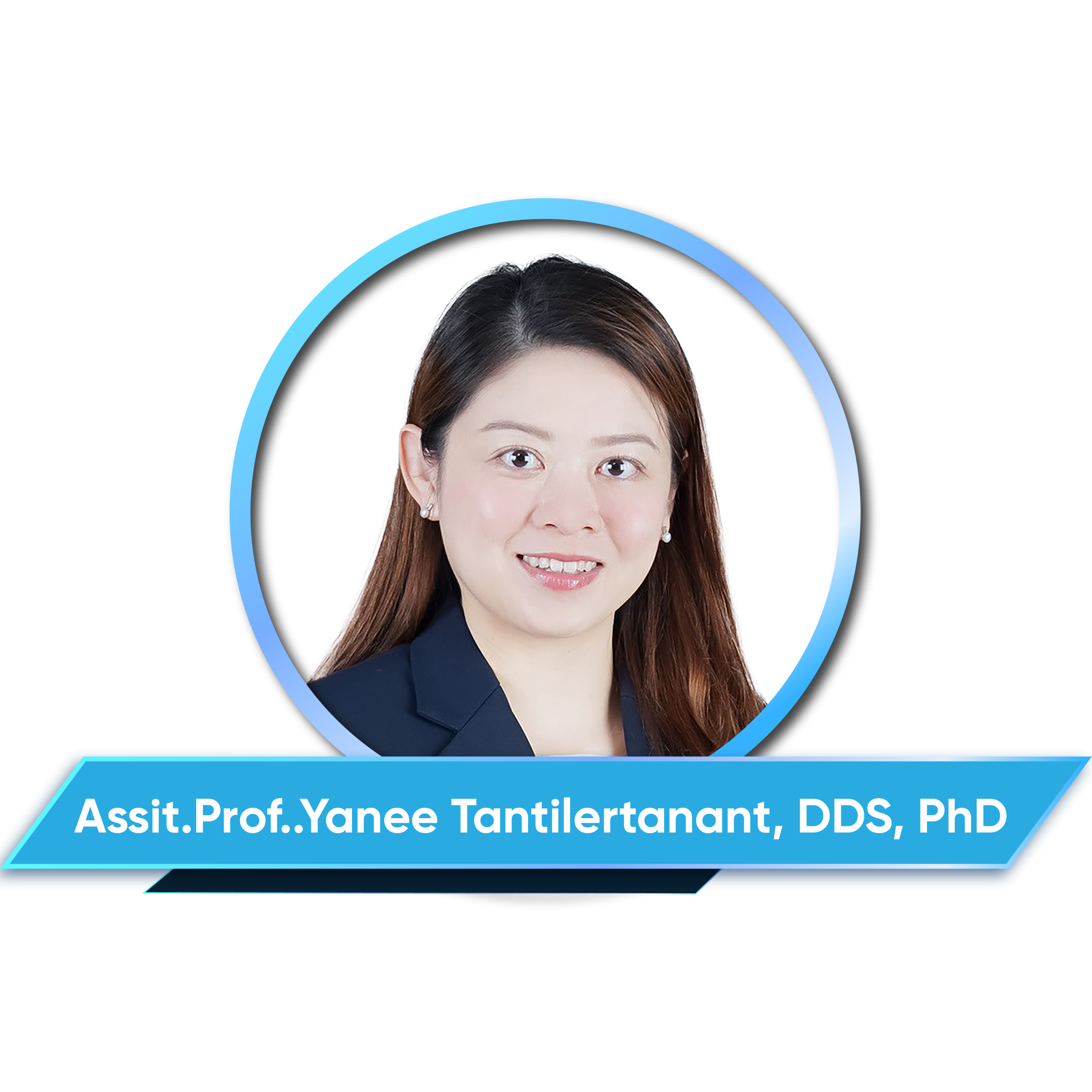
– Received DDS degree from Faculty of Dentistry, Chulalongkorn University, Thailand in 2007.
– Received MSc degree from Faculty of Dentistry, Chulalongkorn University, Thailand in 2013
– Received PhD degree from Faculty of Dentistry, Chulalongkorn University, Thailand in 2019
– Received A.Prof. title from Faculty of Dentistry, Chulalongkorn University in 2024
– Former Instructor at Faculty of Dentistry, Thammasat University, Thailand.
– Instructor at Faculty of Dentistry, Chulalongkorn University
Abstract
Direct resin restoration is a fundamental technique in restorative dentistry, offering a conservative and aesthetic approach to restoring function and form. This procedure relies on composite resins, which provide excellent adhesion, durability, and biomimetic properties when applied with proper protocols. Successful direct resin restorations require an understanding of material selection, preparation techniques, adhesive protocols, layering strategies, and finishing procedures.
Factors such as polymerization shrinkage, shade matching, and moisture control significantly impact the longevity and esthetics of the restoration. This lecture will explore the essential principles of direct resin restorations, highlighting best practices, clinical challenges, and strategies to optimize outcomes. By mastering these fundamentals, clinicians can achieve predictable and long-lasting restorations that enhance both function and patient satisfaction.
CV
– Received DDS degree from Pusan National University, South Korea in 2003.
– Received PhD degree from Pusan National University, South Korea in 2013.
– Received Prof. title from Pusan National University, South Korea in 2014.
– Professor, Dept. of Oral & Maxillofacial Surgery, School of Dentistry, & Dental Hopital, Pusan National University, South Korea.
Abstract
Implants have become an essential dental treatment for functional restoration of the maxillary edentulous molars. However, the presence of the maxillary sinus makes implant placement difficult, and various methods have been suggested to overcome this. However, there are still many controversies, including the surgical method for maxillary sinus augmentation, and the choice of bone grafting materials.
In this lecture, I will show the current status of evidence-based maxillary sinus augmentation through various literature reviews, and I will add my own method to help the audience in their clinical practice.
Objectives:
– Selection of surgical method.
– Selection of graft material.
– Other controversies.
CV
– Received DDS degree from Wonkwang University, Iksan, Chunbuk, South Korea in 1995.
– Received MSc degree in Oral and Maxillofacial Surgery from Korea university Graduate School of Clinical Dentistry, Seoul, South Korea in 2007.
– Adjunct professor, Dept. Of Oral and maxillofacial surgery, Korea university,South Korea.
– Adjunct professor, Dept. Of Oral and maxillofacial surgery, Daegu Catholic University,South Korea.
– Director of international affair, Korean Dental Association.
– Chairman, Dental Public Health Commission of Asia Pacific Dental Federation.
– Director of Seoul top dental hospital.
Abstract
Most dental implants consist of three main components: the fixture, which is osseointegrated into the alveolar bone; the abutment, which connects to the fixture and supports the prosthetic crown; and the crown itself. Generally, the connection between the implant fixture and the crown falls into two categories: screw-retained and cement-retained. A hybrid method known as SCRP (Screw- and Cement-Retained Prosthesis), which combines both approaches, has also been widely used. In screw-retained systems, the crown and abutment are fabricated as a single unit and secured without the use of cement. In contrast, both cement-retained and SCRP systems require an intermediate cementation step to attach the crown to the abutment.
Typically, in the patient’s mouth, the abutment is first connected to the implant fixture, and the crown is then cemented onto the abutment. However, during this process, residual cement may inadvertently remain in the peri-implant area, particularly between the implant and surrounding soft tissue. This is one of the leading causes of peri-implantitis. To address this issue, a variety of cementless implant abutments have been developed in recent years. These designs eliminate the cementation step, thereby reducing the risk of peri-implantitis and simplifying the clinical workflow. Because the crown can be placed and removed directly in the oral cavity, treatment time is also reduced. Although several types of cementless abutments are currently available, they are relatively new and still lack long-term clinical data.
In this report, I will share my clinical experience with cementless abutments, specially their advantages, disadvantages, and potential role in implant prosthetics.
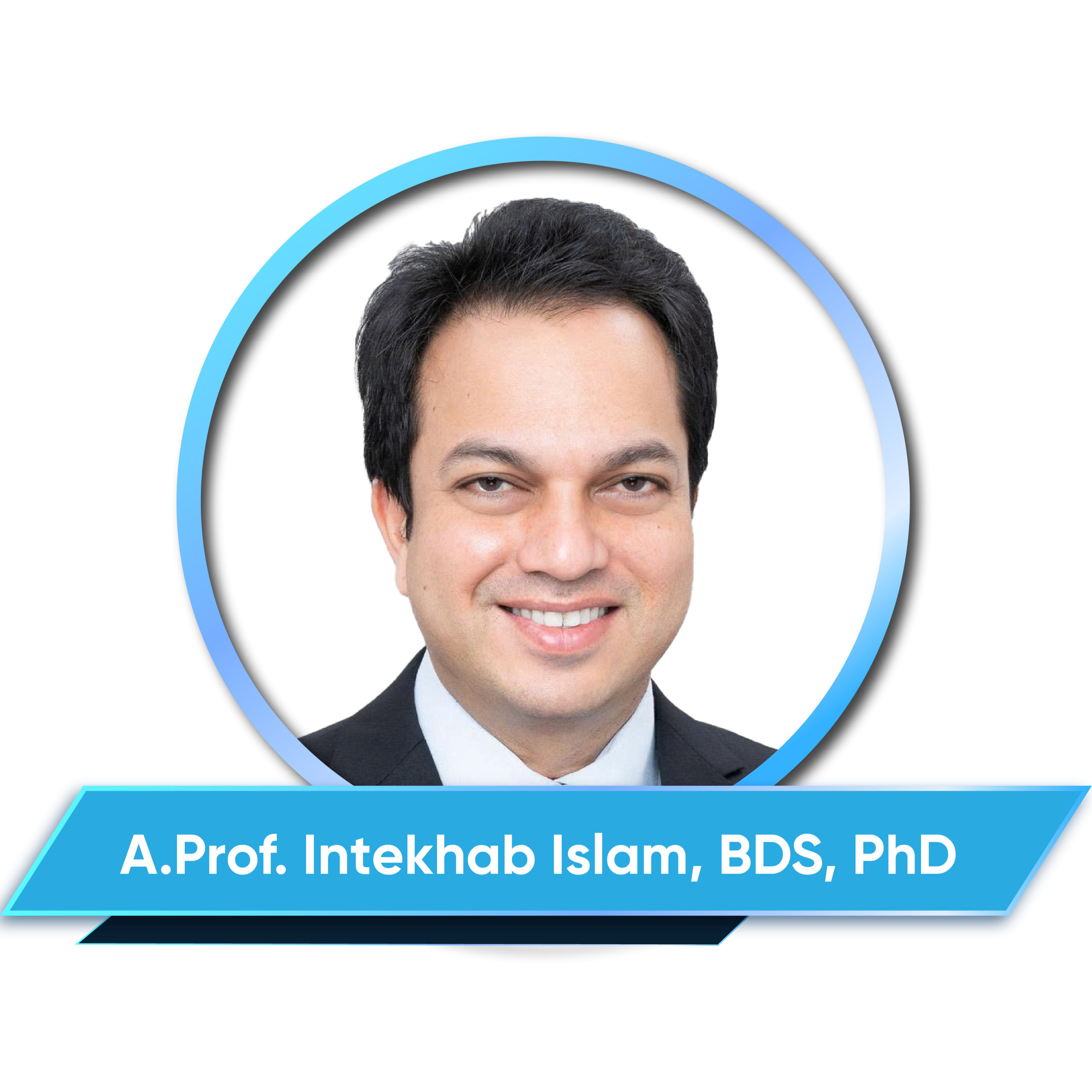
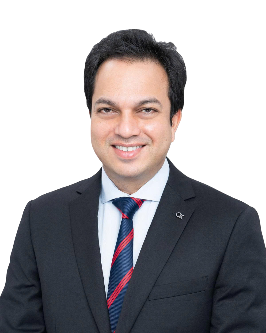
CV
– Receved BDS degree from University of North Bengal, India in 2001.
– Received MSc degree from National University of Singapore in 2005.
– Received MDS degree from National University of Singapore in 2008.
– Fellows of the Academy of Medicine, Singapore in Oral and Maxillofacial Surgery from 2011.
– Received PhD degree from National University of Singapore, Singapore in 2017.
– Chair, Chapter of Oral and Maxillofacial Surgeons, Academy of Medicine, Singapore.
– Chair Total Wellness Safety and Health, Faculty of Dentistry, NUS.
– Chair Clinical Dentistry II Examination Committee, Faculty of Dentistry, NUS.
– Chair Basic Medical Science Education Committee, Faculty of Dentistry, NUS.
– Member, Dental Specialist Accreditation Committee Oral and Maxillofacial Surgery.
– Council Member, Singapore Dental Council.
– Council Member, Singapore Dental Association.
– Associate Professor, Oral and Maxillofacial Surgery, Faculty of Dentistry, NUS.
– Senior Consultant, Oral and Maxillofacial Surgery, NUHS.
Abstract
Antibiotics play a critical role in managing infections in dental practice. However, inappropriate use has contributed significantly to the rise of antimicrobial resistance (AMR), now recognized as a global health threat. This lecture provides an evidence-based overview of when and how antibiotics should be prescribed in dental settings, based on the latest Singapore Clinical Practice Guidelines. It will cover therapeutic and prophylactic use across key specialties including oral surgery, endodontics, periodontology, implantology, paediatric, and geriatric dentistry.
Emphasis will be placed on diagnostic precision, antimicrobial stewardship, and adapting regimens based on patient-specific factors such as systemic health and immunocompromised states. Attendees will gain clarity on clinical scenarios where antibiotics are necessary, discouraged, or require specialist consultation. By aligning with these guidelines, dental practitioners can enhance patient outcomes while contributing to the global effort to combat AMR
Objectives :
– Identify clinical situations in dentistry where antibiotic therapy is indicated versus contraindicated.
– Understand the risks of inappropriate antibiotic use and the mechanisms of antimicrobial resistance.
– Apply principles of antimicrobial stewardship, including drug selection, dosing, and treatment duration.
– Recognize the role of patient-specific factors (e.g., immunocompromised status, age, comorbidities) in guiding antibiotic use.
– Integrate updated evidence-based guidelines into daily dental practice to minimize unnecessary antibiotic prescriptions.
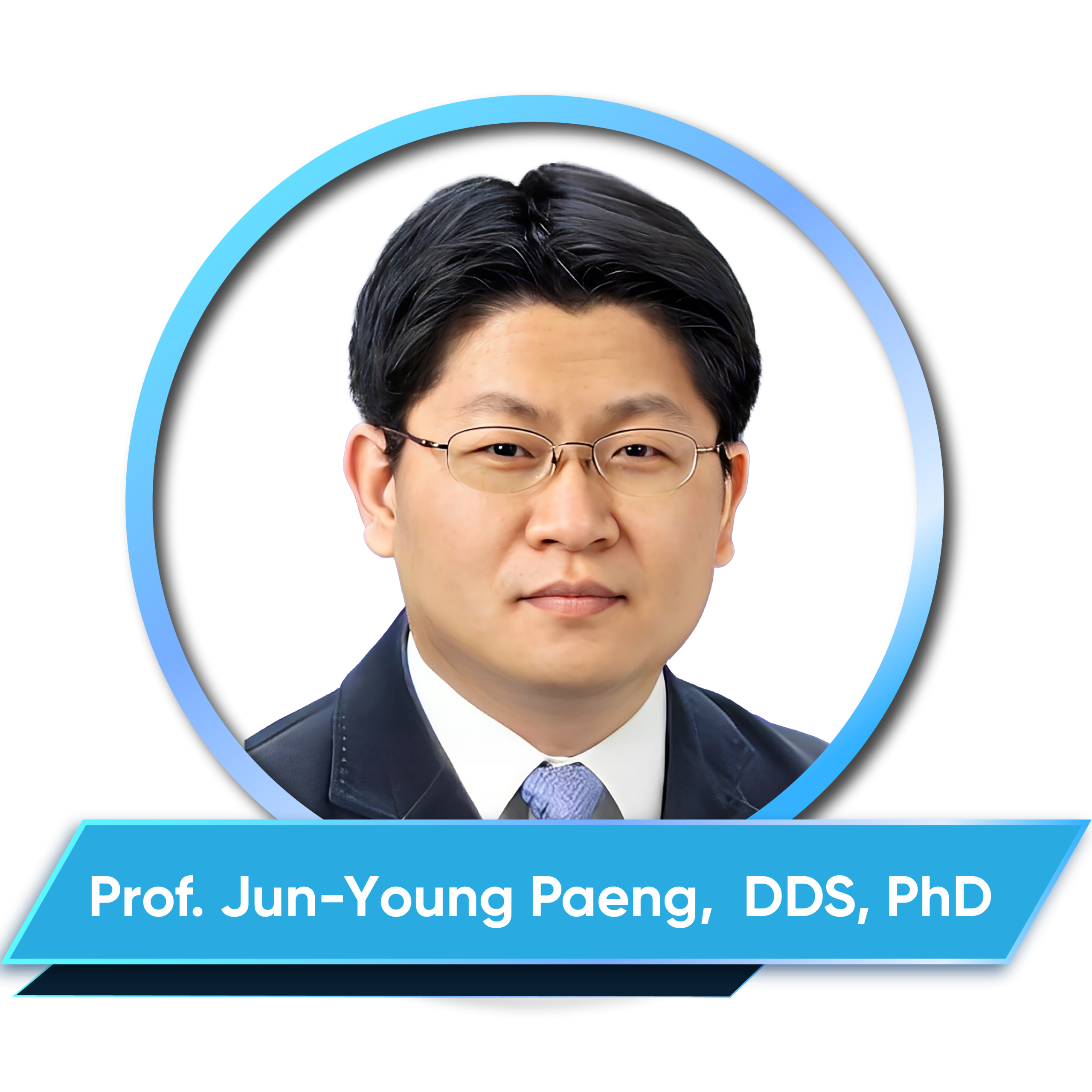
CV
– Received DDS degree from College of Dentistry, Seoul National University, South Korea in 1996.
– Received MSD degree from Graduate School, Seoul National University, South Korea in 1999.
– Received PhD degree from Graduate School, Seoul National University, South Korea in 2005.
– Fellow, Dept. of Oral & Maxillofacial Surgery, Seoul National University Dental Hospital from 2003-2006.
– Visiting Researcher, Oral and Maxillofacial Reconstructive Surgery, Kyushu Dental College, Japan from 2006-2007.
– Assistant Professor, Dept. of Oral and Maxillofacial Surgery, College of Dentistry, Wonkwang University, South Korea from 2007-2010.
– Clinical Associate Professor, Dept. of Oral and Maxillofacial Surgery, Samsung Medical Center, Sungkyunkwan University, Seoul,, South Korea from 2010-2012.
– Assistant professor, Dept. of Oral and Maxillofacial Surgery, Kyungpook National University, Daegu, South Korea from 2013-2018.
– Clinical Professor, Dept. of Oral and Maxillofacial Surgery, Samsung Medical Center, Sungkyunkwan University, Seoul, South Korea from 2018.
Abstract
Alveolar bone reconstruction has been one of the pre-prosthodontic treatment in the field of oral and maxillofacial surgery for a long time. However the term of alveolar bone graft was popularized with the recent advancement of implantology. The implantologist can meet frequently the cases which need alveolar bone augmentation. Sometimes the augmentation procedures show good result, but the number of failure cases increase according to the increased number of the patients.
The advantages of the interpositional bone graft may include followings. The pedicled bone segment shows relatively high resistant to infection and resorption. The crestal bone can be remained on the implant margin and more resistant to marginal bone resorption. The height of the augmentation can be achieved more than GBR to 5-7 mm. The attached keratinized gingiva can be maintained. However, the interpositional bone graft is beneficial when applied to the ridge with adequate alveolar width.
This presentation overviews the limitation of the alveolar bone augmentation, the usefulness and technical point of the interpositional bone graft with literature review.
CV
– Received DDS degree from Osaka Dental University, Japan in 1976.
– Received PhD degree from Osaka Dental University, Japan in 1980.
– Received Prof. title in Esthetic Dentistry from Osaka Dental University, Japan.
– Former President, Nara Dental Association, Japan.
– Standing Director, Japan Dental Association.
– President, Japan Academy of Digital Dentistry
Abstract
In recent years, the use of IT in society has progressed rapidly, and digitalization has progressed in dental treatment.
The digitalization of dental treatment is being promoted along with the introduction of IT in medical information. In particular, the promotion of dental CAD/CAM technology deserves special mention and has revolutionized the concept of the past.The application of dental CAD/CAM technology is effective in improving the diagnostic ability of surgeons, making the practice process more reliable, and improving the environment. This means that patients will be able to provide comfortable, fast, and safe devices.
CV
– Graduated from College of Dental Surgery, Manipal, India.
– Received MSc degrees in Maxillofacial Surgery and also his FDSRCS, FFDRCSI, from the Royal College of Surgeons of England and Ireland respectively.
– Specialist at the Ministry of Health of Malaysia.
– Research investigator for the Oral Cancer Research Cordinating Centre and Cancer Research Malaysia.
– Member of the international faculty of the AO Foundation in Davos Switzerland.
– Mentor and trainer for the Nobel Biocare Implant Training Programme.
– Publication secretary and editor of the Malaysian Dental Journal if the Malaysian Dental Association.
– Past President of the Malaysian Association of Oral & Maxillofacial Surgeons.
– Hon Secretary to the College of Dental Specialist, Academy of Medicine Malaysia.
Abstract
Emergencies in Maxillofacial surgery could be very dramatic anything ranging from torrential bleeding to rip roaring infections or sudden blindness due to complication arising from oral surgical treatment. Emergencies never occur all of a sudden, there is usually progression of the sign and symptoms leading to a full blown emergency. There is a proverbial saying “ What the mind does not know, the eye does not see” What is important to the Oral surgeon and his team is to recognises signs and symptoms well in advance so that necessary precautions and actions taken to prevent such emergencies.
Early intervention in such scenarios reduce the morbidity and mortality outcomes. In this presentation we look at some of the common as well as potential emergencies that we come across in the dental practice settings and we highlight early warning signs and appropriate measures taken to prevent such emergencies. We also look at the current trends in managing such emergencies.
CV
– Received DDS degree with Honors from Indiana University School of Dentistry, USA in 1986.
– Former work positions:
+ Resident Indiana University Hospitals,Hospital Based GPR, Indianapolis, Indiana, USA.
+ Department Head, Deputy Director, Mashoko Hospital, Bikita, Zimbabwe.
+ Country Director / Senior Dental Advisor MOH, Phnom Penh, Cambodia.
– Director / Founder PEACE Dental Clinic, Hanoi, Vietnam.
– Co Founder of SDDS, Smart Dent Delivers Success, Hanoi, Vietnam.
Abstract
Early years orthodontic treatment, providing care for children during the mixed dentition stage, has been a controversial topic. However, today’s dentistry is adding digital diagnostics, digitally designed treatment planning and 3 D printed appliances, employing clear aligners and newer appliances as platforms to effect growth modification and tooth movements. These technical innovations are leading a paradigm shift in orthodontic treatment: proactive early interventions are recommended for optimal development of the young child, from an orthodontic and a wider holistic perspective.
The presenter will address the rationale for mixed dentition treatment, case selection for mixed dentition treatment, and the timing of early interventions. They will share from their years of experience with classical functional appliances and 5 years of experience with clear aligners providing mixed dentition orthodontic therapy. The aim of this session is to effect a change in your practice paradigm and open for you, doctor, a new and very beneficial area of treatment.
CV
– Received DDS degree from Renn University, France in 1997.
– Received Residency doctor degree in maxillofacial surgery from Lille University, France in 2000.
– Received PhD degree from Hanoi Medical University in 2006.
– Received Associate Professor title in 2015.
– President of Council of the University of Medicine and Pharmacy, Vietnam National University, Hanoi.
– Head of Orthodontic Department, School of Medicine and Pharmacy, Vietnam National University, Hanoi.
– Member of the executive committee, Vietnam Association of Oral Maxillofacial and Plastic Surgery.
– Member of the executive committee, Vietnam Association of Orthodontists.
– Member of World Federation of Orthodontists.
Abstract
Nonsurgical treatment of Class II malocclusion depends on the skeletal or dental cause, age, and severity of the deviation. In growing children, the goal is to correct skeletal growth with functional appliances with or without Headgear to limit maxillary growth. In adults, treatment tends to involve camouflage by extraction of upper premolars, use of mini screws to distalize the maxillary dental mass, or rotation of the incisor axis. Cephalometric and soft tissue analysis help determine the appropriate treatment approach. Profile esthetics, occlusal function, and patient compliance should be evaluated. Treatment should be individualized and incorporate maintenance strategies to ensure longterm, stable results.
CV
– Received DDS degree from HCMC University of Medicine and Pharmacy in 1997.
– Received PhD degree from Paris V University, France in 2013.
– Received Msc degree from Paris V University, France in 2007.
– Received Certificate in Dental Biomaterials from Paris V University in France in 2004.
– Received Certificate in Dental Implantology from Paris VI University in France in 2012.
– Received Assoc. Prof. title in 2022.
– Head of Department of Dental Basic Sciences, HCMC University of Medicine and Pharmacy.
Abstract
The art of layering has long been used to create natural-looking ceramic teeth. Traditionally, feldspathic ceramics, with a vitreous matrix and fine crystalline filler, were employed. However, recent decades have seen major advancements in dental ceramics, notably zirconia and lithium disilicate ceramics.
Zirconia, valued for its toughness and flexural strength, enables the construction of extensive tooth- or implant-supported frameworks, effectively replacing traditional metallurgical structures. Lithium disilicates, with a vitreous matrix and large crystalline filler, offer notable durability and are ideal for veneers due to their excellent bonding and aesthetic properties.
This lecture will discuss appropriate indications, new concepts, and clinical protocols for dentin adhesion, with a particular focus on zirconia veneers.
CV
– Received DDS degree from HCMC University of Medicine and Pharmacy in 1990.
– Received MSc degree of Medicine from HCMC University of Medicine and Pharmacy in 1998.
– Received PhD degree in Maxillofacial Surgery from 108 Institute of Clinical Medical and Pharmaceutical Sciences in 2011.
– Received MSc degree of Dentistry, specialized in Orthodontics, Munster University, Germany.
– Vice Dean, Faculty of Dentitry, Head of Department of Oral Pathology and Maxillofacial Surgery, Pham Ngoc Thach Medical University from 2017-2019.
– Dean, Faculty of Dentitry, Head of Department of Oral Pathology and Maxillofacial Surgery, Head of Department of Orthodontics and Pedodontics, Chairman of the Ethics Committee in Biomedical Research, Van Lang University.
Abstract
Orthodontics is a treatment that changes the entire occlusion. Changing the occlusion will risk causing occlusal and temporomandibular joint problems. Orthodontic patients may have a healthy temporomandibular joint or a temporomandibular joint disorders acompany. The temporomandibular joint disorder may worsen after orthodontic treatment if the principles of restoring optimal functional occlusion are not followed. Orthodontic treatment can also be applied to treat temporomandibular joint disorders and in this case, it may be necessary to change the position of the condyle before orthodontic treatment, to ensure the reestablishment and stabilization of the optimal condyle position.
Ensuring respect for occlusal references is the most important principle in orthodontic treatment, especially in patients with temporomandibular joint disorders.
CV
– Received DDS degree from HCMC University of Medicine and Pharmacy in 1997.
– Received Resident physician degree from HCMC University of Medicine and Pharmacy in 2002.
– Received PhD degree from 108 Institute of Clinical Medical & Pharmaceutical Sciences in 2015.
– Chairman of Nhan Tam Dental Clinic.
– Head of Dental Implant Division, Hong Bang International University.
Abstract
Dental implants are recognized as a standard treatment for restoring tooth loss; however, challenges persist regarding implant precision, complications, and inconsistent outcomes, primarily due to the surgeon’s experience. Artificial Intelligence (AI) and Robotics have introduced a transformative approach: AI analyzes 3D imaging to optimize implant positioning, while Robotic systems enable minimally invasive surgery with submillimeter accuracy and anatomically guided drill control. This technology enhances success rates, improves safety, and facilitates treatment personalization. However, limitations include high implementation costs, specialized training requirements, and risks of technical failure. This presentation outlines the revolutionary role, clinical applications, and transformative potential of AI/Robotics in dental implantology.
Objectives:
– Define and differentiate between AI and Robotics.
– Understand AI applications in dental implantology.
– Comprehend the role of AI/Robotics in dental implant surgery.
CV
– Received DDS degree from Hanoi Medical University in 1997.
– Received MSc degree from Hanoi Medical University in 2005.
– Received PhD degree from 108 Institute of Clinical Medical and Pharmaceutical Sciences in 2014.
– Received the Certificate of Fundamentals of Endodontics from Boston University in 1998, Certificate of Advanced Endodontics from Alberta University in 1998.
– Received the Certificate of Microscopic Endodontics from Chulalongkorn University, Thailand in 2014.
– Received the Certificate of Endodontic Microsurgery from Pennsylvania University, USA in 2024.
– Head of High- Technology Dental Department, National Hospital of Odonto- Stomatology, Hanoi.
Abstract
Vital pulp therapy (VPT) has emerged as a cornerstone of minimally invasive endodontics, offering a paradigm shift in the management of irreversible pulpitis. This presentation explores the full spectrum of VPT’s potential, emphasizing its application in preserving pulp vitality even in cases traditionally deemed for root canal treatment. A deep dive into the nuances of diagnosis will be provided, highlighting the importance of correlating clinical findings with advanced understanding of pulp histopathology and histobacteriology. The interplay between pulpal inflammation, tissue healing, and bacterial control will be discussed, underlining the biological basis for successful outcomes.
Special focus will be given to the pivotal role of calcium silicate cements, whose bioactivity and sealing ability have revolutionized VPT procedures. Through evidence-based scenarios and clinical insights, this presentation aims to equip clinicians with the knowledge to accurately diagnose, select cases, and apply VPT techniques effectively. Attendees will gain a comprehensive understanding of how to harness VPT to achieve predictable, long-term success in the management of irreversible pulpitis.
CV
– Received DDS degree from Faculty of Dentistry, HCMC University of Medicine and Pharmacy in 2002.
– Received PhD degree from Aix-Marseille University, France in 2008.
– Vice-Dean of the Faculty of Dentistry, Van Lang University.
– President of the Ho Chi Minh City Society of Dental Implantology.
– President of ITI Vietnam (International Team for Implantology – Vietnam Section).
Abstract
The integration of digital workflows into contemporary implant treatment has significantly transformed clinical practices by enhancing accuracy, reproducibility, and efficiency. This integration can range from partial to fully digital workflows, aiming to streamline and optimize the treatment process—from creating digital replicas of patients to treatment planning, guided surgery, and prosthetic fabrication. The incorporation of artificial intelligence further refines treatment planning by predicting optimal implant positions and potential complications. Digital workflows promote interdisciplinary collaboration, enhancing communication between doctors and technicians, which leads to more cohesive treatment plans.
This modern approach not only elevates the standard of care but also meets the growing demand from patients for minimally invasive, efficient, and highly customized dental solutions. This presentation focuses on analyzing the applications of digital workflow in contemporary implant treatment through a literature review and a variety of clinical case scenarios.
Objective: Analysis of the advantages of digital procedures in contemporary implant treatment.
Topic 2: Applications of 3D technology in maxillofacial surgery: A paradigm shift
CV
– Received DDS degree from Hanoi Medical University in 2011.
– Received MSc degree in Oral and Maxillofacial Surgery from The University of Manchester, United Kingdom in 2015.
– Received PhD degree in Medical Research from Shimane University, Japan in 2021.
– Clinical Fellowship in Oral and Maxillofacial Surgery in Department of Oral and Maxillofacial Surgery, Shimane University Faculty of Medicine from 2016 to 2021
– Clinical Fellowship in Oral and Maxillofacial Surgery (Minimally invasive Orthognathic surgery) in AZ Sint-Jan Hospital, Belgium in 2023
– Clinical Fellowship in Craniofacial Surgery in Heidelberg University Hospital, Germany in 2025
– Oral and Maxillofacial surgeon, Dept. of Plastic and Aesthetic Surgery, National Hospital of Odonto-Stomatology, Hanoi.
– Vice President, Vietnam 3D Technology in Medicine Association.
Abstract
Abstract 1: In-depth research into mutated genes causing ameloblastoma is ushering in a new era for treating this condition. Previously, the primary method was often surgical removal of part or all of the jawbone, significantly impacting patients’ quality of life. However, identifying specific gene mutations, particularly BRAF V600E and SMO, has laid the groundwork for the development of targeted therapy. This therapy directly targets abnormal proteins produced by the mutated genes, inhibiting tumor growth and spread without harming healthy cells. As a result, many ameloblastoma patients can now avoid complex jawbone surgeries, minimizing complications and preserving anatomical structure and function for chewing and speaking. This…
Abstract 2: The 3D technology has gradually played an important role in contemporary oral and maxillofacial surgery. The value of this technology is the fact that it not only improves the accuracy of the operation but also makes the complex procedures feasible. From post-ablation reconstruction with free tissue transfer, orthognathic surgery to panfacial trauma, all of these can be assisted to simplify intraoperative protocols. The 3D technology helps surgeons evaluate pre-surgical anatomy and design surgical guide to make the final outcome to closely resemble the treatment plan. In this presentation, some complicated cases in maxillofacial surgery will be presented.
Topic 2: Freehand or digital workflow in full-arch implant rehabilitation: Which approach offers superior clinical outcomes?
CV
– Received DDS degree from Hanoi Medical University in 1999.
– Received PhD degree from Hanoi Medical University in 2013.
– Head, Department of Oral Implantology, National Hospital of Odonto-Stomatology, Hanoi.
– Lecturer at Department of Implantology, School of Dentistry, Hanoi Medical University.
Abstract
Full-arch implant-supported prosthesis has become an increasingly popular solution for the treatment of complete edentulism. However, the choice between the freehand technique (implant placement without a surgical guide) and the guided digital workflow (with surgical guide) remains inconclusive. This report aims to clarify the clinical differences between these two approaches, focusing on aspects such as accuracy, surgical time, immediate loading capability, complications, and patient satisfaction.
Objectives:
– Clarify the roles and clinical indications of each approach (freehand and digital-guided) across a variety of treatment scenarios, from simple to complex cases.
– Propose a practical direction for the integration of digital workflows in full-arch implant surgery and prosthodontics, tailored to the actual conditions of the clinical setting.
CV
– Received DDS degree from University of Medicine and Pharmacy, Ho Chi Minh City, Vietnam in 1994.
– Received Master of Science in Periodontology from International Medical College, Munster, Germany in 2016.
– Received Certificate of Master Clinician in Implant Dentistry from gIDE institute and Loma Linda University, Los Angeles, USA in 2018.
– Lecturer, Head of Periodontics and Implant Dentistry Department, Faculty of Dentistry, Van Lang University, Ho Chi Minh City, Vietnam.
Abstract
The most challenging goal of periodonttitis treatment is to regenerate the structural components of the damaged periodontal tissue, restoring function and aesthetics to diseased teeth. Scientific research in recent years has demonstrated the ability of self-regenerate of periodontal tissue after treatment and at the same time clarified the role of factors affecting the success of periodontal regenerative treatment.
Nowadays, periodontal tissue regeneration can become a reality with basic periodontal treatments such as non-surgical or complex ones such as advanced periodontal surgery, depending on the clinical situation. The presentation will analyze the factors that determine the ability of periodontal tissue regeneration in the treatment of periodontitis. Indications and treatment methods for each specific case will be mentioned and illustrated with images of real clinical cases.
Objectives:
– Analysis of factors determining the regeneration ability of periodontal tissue in the treatment of periodontitis.
– Selecting regenerative periodontal treatment procedure for each clinical case.
– Received DDS degree from Taipei medical University in 1995 – Received PhD degree from Nihon University ,Japan in 2004 – Received Prof. title from Taipei Medical University in 2016 – Former work positions: President, Taiwan Association of Dental Sciences – Current work positions: Vice President, APDF/ APRO
CV
ABSTRACT
– Received DDS degree from the University of Santiago de Compostela, Spain. – Postgraduate training in Periodontics and Implantology in Madrid; Occlusion and Temporo-mandibular Dysfunction in Valencia and Orthodontics in University of Santiago de Compostela, Spain. – Director of Post-graduate program in Implants and Implant Prosthesis, UAX University, Madrid, Spain. – Adjunct Assistant Professor of the Department of Prosthodontics at LSUHSC School of Dentistry, New Orleans, Louisiana, USA. – Founder of MEDA Training Center, Spain.
Participants will understand principles of vertical ridge augmentation, indications and limitations, case selection, treatment planning, surgical techniques for flap advancement & primary closure, step-by-step surgical protocol, membrane selection, fixation devices, bone harvesting regions and techniques, postoperative protocol, and simultaneous implant placement.
CV
ABSTRACT
– Nhận bằng Bác sĩ nha khoa tại Trường Nha khoa – Đại học Wonkwang, Iksan, Chunbuk, Hàn Quốc năm 1995. – Năm 2007, nhận bằng Thạc sĩ khoa học (MSc) khoa Phẫu thuật hàm mặt của hệ sau đại học của Đại học Hàn Quốc, khoa Nha khoa Lâm sàng, tại Seoul, Hàn Quốc – Giáo sư kiêm nhiệm, khoa Nha khoa, trường Y khoa Công giáo, Đại học Hallym, Đại học Hàn Quốc – Chủ tịch Bệnh viện Nha khoa Seoul Top – Giám đốc Quan hệ Quốc tế của Hiệp hội Nha khoa Hàn Quốc năm thứ 30 – Nghiên cứu viên cao cấp của Viện nghiên cứu implant Hàn Quốc – Chủ tịch danh dự của Học viện Thiên niên kỷ mới – Phó chủ tịch Hội nghị quốc tế về cấy ghép nha khoa (ICOI) – Hàn Quốc – Giám đốc Học viện Cấy ghép răng miệng và hàm mặt Hàn Quốc (KAOMI) – Giám đốc Học viện Nha khoa Thẩm mỹ Hàn Quốc (KAED) – Phó chủ tịch Học viện Nha khoa Laser Hàn Quốc – Thành viên tích cực của Học viện Tích hợp xương (AO) – Trung tâm Giáo dục và Nghiên cứu Cấy ghép Nha khoa của Osstem (AIC) – Giảng dạy tại Viện Nha khoa Laser Thế giới (WCLI) – Chủ tịch Ủy ban Nha khoa cộng đồng APDF/APRO
Định nghĩa về mini-implant thường bị nhầm lẫn với nhiều tên gọi khác nhau vì cụm từ này vừa dùng để chỉ implant trong nha khoa với độ rộng nhỏ, vừa dùng cho TAD (khí cụ neo giữ tạm thời) được sử dụng tạm thời để neo chặn trong chỉnh nha. Hơn nữa, trước đây, hầu hết implant có chiều rộng dưới 3,0 mm được chế tạo dưới dạng implant một khối vì trong trường hợp trụ implant được kết nối với khớp nối Abutment, thành implant mỏng có thể bị vỡ do lực nhai. Tác giả này cho rằng mini implant hẹp nên được định nghĩa chính xác là một implant có chiều rộng dưới 3,0mm được sử dụng để chữa trị những răng bị mất, nhưng do sự ra đời gần đây của implant có kết nối với răng mài bằng vít nhỏ hơn 3,0mm, việc xác định mini implant hẹp theo độ rộng nhất định hay hình dạng implant một khối gặp khó khăn. Trong trường hợp mất răng ở những vùng có bề rộng xương ổ răng nhỏ như răng trước hàm dưới thì việc lựa chọn vật liệu cố định implant và phương pháp phẫu thuật là rất khó khăn, cấy ghép implant với chiều rộng và phương pháp thông thường có thể gây ra các vấn đề như implant thất bại, hoặc làm tổn thương các răng lân cận. Để tránh những biến chứng này, các kế hoạch điều trị khác nhau có thể được thực hiện tùy thuộc vào số lượng và vị trí của răng bị mất, cũng như lựa chọn của bác sĩ phẫu thuật, nhưng tác giả này cho rằng việc sử dụng mini implant hẹp một khối sẽ là một lựa chọn tốt trong trường hợp chiều rộng cơ tim hẹp. Tại đây, ông có báo cáo rằng ông đã quan sát thấy hiệu quả lâm sàng tốt của mini implant hẹp một khối trong thử nghiệm lâm sàng của mình và nghĩ rằng nếu phẫu thuật được thực hiện bằng cách sử dụng chỉ dẫn bằng kỹ thuật số để lắp đặt chính xác, nó sẽ hướng đến phương pháp điều trị chính xác và đáng tin cậy hơn
CV
ABSTRACT
– Sáng lập công ty Dentium và công ty Genoss Hàn Quốc năm 2000 – Giáo sư khoa Nha chu, Đại học Kyung Hee Hàn Quốc từ năm 1993 – Giáo sư Nha khoa, ĐH Korea từ năm 1993 – Phó giáo sư và nhà nghiên cứu lâm sàng Trung tâm Implant, Đại học Tuebingen, Đức từ năm 1999 – 2000 – Nghiên cứu viên, Giáo sư tại Viện nghiên cứu, Trung tâm Implant, Đại học Loma Linda, Hoa kỳ từ năm 2000 – 2001. – Giám đốc phụ trách chuyên môn Nha khoa, Quốc tế Well – Seoul, Hàn Quốc từ năm 2001.
Updating…
CV
ABSTRACT
– Received BDS degree from University of Malaysia in 2008. – Member of the Faculty of Dental Surgery of The Royal College of Surgeons of Edinburgh (MFDS RCSEd) since 2014. – Earned a specialist degree in Periodontology (MClinDent) from University of Malaya in 2017. – Periodontist at the Kota Setar Specialist Clinic, in Kedah state of Malaysia. – Member of Malaysian Society of Periodontology(MSP), Malaysian Dental Association (MDA) and International Team for Implantology (ITI).
Mucogingival deformities and gingival recession are a group of conditions that affect a large number of patients. There is an increased risk of development or progression of gingival recession in cases presenting with thin periodontal phenotypes, suboptimal oral hygiene, and requiring restorative/ orthodontic treatment. Aesthetic concern, dentin hypersensitivity, cervical lesions, thin gingival phenotypes and mucogingival deformities are best addressed by mucogingival surgical intervention when deemed necessary. Learning Objective: – Review terminology of mucogingival complex, classification of gingiva recession, periodontal and perimplant phenotype. – Learn how to identify the mucogingival deformities and conditions. – Recipient and donor site preparation and harvesting techniques. – Review common periodontal plastic surgery procedures focusing on periodontal phenotype modification therapy, rationale and indications for surgery, alternative options and key takeaways from the treatments.
Lecture 1: Benefit and Significance of Deep Shape in Endodontics: Illustrated with Clinical Case Discussion
CV
ABSTRACT
– Received BDS degree from University of Pretoria, South Africa in 1992. – Received Diploma in Endodontics (Cum Laude), Masters Degree in Endodontics (Cum Laude) and a Diploma in Aesthetic Dentistry (Cum Laude) and PhD degree in Endodontics. – Professor in the Department of Odontology, School of Dentistry, University of Pretoria, South Africa. – Past President of the South African Academy of Aesthetic Dentistry (SAAAD). – President of The South African Society for Endodontics and Aesthetic Dentistry (SASEAD).
Deep Shape is necessary to ensure excellent mechanical debridement of the apical third of root canal system, while maintaining the apical extent of the root canal as small as practical with sufficient taper behind the physiologic terminus. It further allows for the safe exchange and activation of irrigant to ensure 3D disinfection and predictable canal obturation to prevent reinfection. It is important that modern endodontic file systems offer a comprehensive set of files with different sizes and tapers, allowing clinicians to adapt to various canal anatomies and to create deep shapes. Incorporating advance metallurgy enhances the flexibility, resistance to cyclic fatigue, and cutting efficiency of the files. These features can contribute to the system’s ability to shape small, large, oval and curved canals, reduce the risk of instrument fracture, and improve overall treatment outcomes. Modern file system should also help to manage access cases that present with open apices and require the placement of MTA. By incorporating these concepts into clinical practice and integrating proper irrigation and obturation techniques, partitioners can enhance the overall quality of root canal therapy, leading to improved treatment outcomes for their patients. Learning objectives: – Review the goals of endodontics. – Discuss the concept of deep shape in endodontics. – Review the advantages of deep shape during canal preparation, irrigation and obturation. – List the advantages of a contemporary file systems. – Demonstrate the clinical application of deep shape in clinical case reports.
Lecture 2: Restorative Protocols for Endodontically Treated Teeth
CV
ABSTRACT
– Received BDS degree from University of Pretoria, South Africa in 1992. – Received Diploma in Endodontics (Cum Laude), Masters Degree in Endodontics (Cum Laude) and a Diploma in Aesthetic Dentistry (Cum Laude) and PhD degree in Endodontics. – Member of SADA, IADR, NOMADS, Study Group 1, Endodontic Society of South Africa, Dental Materials Group and the Academy for Microscope Enhanced Dentistry (AMED). – Professor in the Department of Odontology, School of Dentistry, University of Pretoria, South Africa. – Past President of the South African Academy of Aesthetic Dentistry (SAAAD). – President of The South African Society for Endodontics and Aesthetic Dentistry (SASEAD).
This presentation will address with the “Root 2 Crown” concept that will enable clinicians to provide a high level of care to their patients after Endodontic treatment. It is a well-established fact that endodontic success can drop from 90% to 18% if the coronal restoration is poor or delayed. Concepts that will be discussed include pre-endodontic restorations, modern endodontic principles regarding root canal preparation, differences between vital and non-vital teeth, guidelines for restoration of Class I, Class II and more severely broken-down teeth after endodontic treatment. Guidelines for the placement of posts will also be reviewed. The restorative concepts and materials discussed will enable clinician to achieve more predictable long-term results in clinical practice. The aim and objectives of this presentation: Understand the importance of coronal seal after endodontic treatment. Learn how to restore Class I and Class II access cavities predictably. Realize the benefit of using fiber posts. Understand when and when not to place a post. Understand when to use direct or indirect restorations.
CV
ABSTRACT
– Nhà nghiên cứu và lâm sàng nổi tiếng thế giới về Nha khoa can thiệp tối thiểu và bệnh lý sâu răng – Giáo sư đại học Queensland, Australia – Nguyên Trưởng khoa Nha, Đại học Kuwait, Đại học Sharjah, UAE – Giáo sư thỉnh giảng của: + King College, London + Đại học Queensland, Australia + Đại học Malaya, Kuala Lumpur, Malaysia + Đại học Singapore, Singapore Đại học Thammasat, Bangkok, Thailand
Despite continuing major advances in dental materials and techniques. The average longevity of a direct tooth coloured restoration is still hovering around 10 years. Restorative materials are still poor substitute for natural tooth structure. Teeth can withstand high mastication load because they are built using two very different materials, so it has been suggested that we should also replicate this design when rebuilding a tooth. Today, technological innovations have provided dental professionals with new tools and science has provided us with many possible ways of handling the above issues. This lecture aims at identifying important factors that govern clinical success, reviewing possible solutions and demonstrating practical ways at preserving and restoring tooth structure. Learning objectives: – Understanding the role of Glass Ionomer Cements in restorative dentistry – How to maximise the clinical performance of Glass Ionomer Cements and Composite Resin – Conservative restorative techniques, alternative options for restoration, and maintaining restored dentitions – Understanding appropriate materials selection and gaining maximum benefits
CV
ABSTRACT
– Received DDS degree from Ministry of Health, Labour and Welfare in 1995. – Received PhD degree from Graduate School, Yokohama City University (YCU) in 2002. – Received Prof. title from YCU Hospital in 2020. – Former Research Associate, Stanford University, USA in 2006. – Former Assistant Professor, YCU Graduate School of Medicine in 2010. – Former Associate Professor, YCU Graduate School of Medicine in 2013. – Deputy director in Dept of Oral and Maxillofacial Surgery in YCUH. – Councilor, Japanese Society of Oral and Maxillofacial Surgeons. – Dental Clinical Instructor for Foreign Dental Practitioner. – Specialist and Instructor of Board Certification in JSOMS. – General Clinical Oncologist in Oral & Maxillofacial Surgery. – Fellow of International Board for Certification of Specialists in Oral & Maxillofacial Surgery (FIBCSOMS)
The UICC and WHO define oral cancer as cancer arising in the buccal mucosa, upper and lower gingiva, hard palate, tongue, and oral floor, and propose a TNM classification based on the size of the primary tumor, regional lymph node metastasis, and the presence of distant organ metastasis. Histopathologically, more than 90% of oral cancers are squamous cell carcinomas. The standard treatment of oral cancer is generally surgery in many cases, and recent advances in multimodality therapies using combination of chemotherapy, radiotherapy, and surgery lead to expanding its indication. However, advanced oral cancer is deadly and disfiguring disease for which better systemic therapy is enormously sought to improve the mortality and avoiding dysfunction. In this talk, I would like to offer commentary on the global standards for the types, characteristics, diagnosis, and treatment of oral cancer. Besides, prevention of oral cancer including our approach for cancer screening would be introduced.
CV
ABSTRACT
– Received DDS degree from Faculty of Dentistry University of Medicine and Pharmacy at Ho Chi Minh City in 2002. – Received PhD degree from Faculty of Dentistry, Marseille, French Republic in 2008. – Former work position : Lecturer at the Department of Prosthodontics, Faculty of Odonto-Stomatology in HCM; – Current work positions: Co-founder of Elite Dental Group ; Vice Dean of Research and International Affairs at Faculty of Dentistry, Van Lang University ; ITI Fellow, President of Ho Chi Minh city society of Dental Implantology (HSDI).
Update...
CV
ABSTRACT
⁻ Nhận bằng BS RHM tại Trường ĐH Y Hà Nội năm 2000 ⁻ Nhận bằng TS RHM tại Trường ĐH Gifu, Nhật Bản năm 2009 ⁻ Vị trí công tác hiện tại: Phó giám đốc CM; Trưởng khoa Răng Miệng, Phó Giám đốc Bệnh viện Hữu Nghị Việt Nam – Cu Ba
Lấy dấu chính xác vị trí implant và chế tác phục hình khít sát thụ động là yếu tố quan trọng đảm bảo sự thành công lâu dài của phục hình toàn hàm trên implant. Quy trình kỹ thuật số hiện nay giúp tối ưu hóa thực hành implant toàn hàm bằng các công cụ lấy dấu chính xác vị trí implant, mô mềm và chế tác phục hình tức thì cho bệnh nhân. Đồng thời việc chuyển đổi từ phục hình tạm sang phục hình sau cùng cũng được đảm bảo về chức năng; khớp cắn và thẩm mỹ. Bài báo cáo sẽ trình bày các bước phẫu thuật với máng đa tầng; quy trình lấy dấu tức thì phối hợp các công cụ lấy dấu chính xác như máy quét trong miệng; quét khuôn mặt; quang trắc lập thể máy PIC; và các kinh nghiệm lâm sàng để nâng cao độ chính xác khi lấy dấu kỹ thuật số; phương thức tương tác với labo để chế tác phục hình tức thì sau phẫu thuật và chuyển đổi sang phục hình sau cùng bằng các quy trình số với vật liệu phù hợp nhất.
CV
ABSTRACT
⁻ Nhận bằng BS RHM tại Trường Đại học Y Dược TP. HCM năm 2005 ⁻ Nhận bằng ThS Khoa học sức khỏe tại Đại học Bordeaux 2, Pháp năm 2007 ⁻ Nhận bằng TS RHM (Sinh học miệng) tại Trường Mahidol, Thái Lan năm 2013 ⁻ Nhận chức danh PGS năm 2022 ⁻ Các vị trí công tác đã trải qua: Trưởng bộ môn Nha chu, Phó trưởng khoa RHM Trường Đại học Y Dược TP. HCM ⁻ Vị trí công tác hiện tại: Trưởng bộ môn Nha chu, Phó trưởng khoa RHM Trường Đại học Y Dược TP. HCM
Bệnh nha chu là một tình trạng răng miệng rất phổ biến ảnh hưởng đến các mô nâng đỡ răng. Bằng chứng khoa học đang được tích lũy về mối liên hệ giữa bệnh nha chu và các tình trạng toàn thân khác nhau. Bài tổng quan này cung cấp góc nhìn tổng quan toàn diện, cập nhật và ngắn gọn về chủ đề này, đặc biệt kết hợp các kết quả nghiên cứu thực hiện tại Việt Nam, hy vọng hỗ trợ các nhà thực hành Răng Hàm Mặt và Y khoa cung cấp dịch vụ chăm sóc tích hợp để có kết quả lâm sàng tốt hơn. – Giải thích được cơ chế đề nghị về mối liên quan giữa bệnh nha chu và sức khỏe toàn thân dựa trên thực chứng – Phân tích được mối liên quan giữa bệnh nha chu và một số bệnh toàn thân – Ứng dụng được trong chẩn đoán và điều trị Răng Hàm Mặt và Y khoa
CV
ABSTRACT
Dental implant has been used in the world for 50 years and became a popular therapy in the dentistry. However, a lot of problems were occurred with the dental implant therapy. First, systemic disease problems are the most common problem caused to the implant surgical complication. Second, bone quality and quantity are the important consideration for the implant placement. Therefore, hard tissue management, which is the most complicated and difficult, is always a key factor to lead the implant success. On the other hand, the management of the soft tissue around the dental implant, which were influenced the complication of the dental implant, will be discussed. Besides, the peri-implantitis also usually occurred in the dental implant therapy. The etiology and treatment of peri-implantitis are the critical issue in the implant maintenance. Finally, the mechanical problem analysis and management, a serious and interesting issue, will be investigated for the physical success.
CV
ABSTRACT
– Received DDS degree from Intercontinental University in 1995 – Received Master of Science degree from Loma Linda University in 1998 – Received “Professor Honoris Causa” title from – Former work positions: Invited Professor Zaporizhzhia State University, Ukraine, 2021 – Current work positions: Assistant Full-time Professor of Loma Linda University, Orthodontic Department
In the vertical growth pattern, the dentoalveolar symptoms are protrusion of the upper incisors and a lingual inclination of the lower incisors. In the horizontal growth pattern, the dentoalveolar symptoms are proclination of the upper and lower incisors. In the Skeletal open bite, the anterior facial height is excessive, especially at the level of the lower third, while the posterior height (branch height) is short. Treatment should be aimed at intruding the extruded upper teeth or preventing them from erupting at the same time as the other structures grow (relative intrusion). Elongated face syndrome. Learning objectives: The Audience will learn how to 1) Diagnose, and Establish, the Orthopaedic, Orthodontic,and Surgical Correction of Open bites
CV
ABSTRACT
– Received DDS degree from University of Chieti, Italy in 2001. – Received Certificate in Orofacial Pain from University of Kentucky, USA in 2007. – Received MS degree in Orthodontics from University of Cagliari, Italy in 2012. – Former Clinical Associate Professor at the University of North Carolina at Chapel Hill. – Clinical Associate Professor at Indiana University School of Dentistry.
Temporomandibular disorders (TMD) represent a diverse group of conditions affecting the temporomandibular joints (TMJ), masticatory muscles, and associated structures. These disorders manifest as pain, TMJ sounds, and limitations in jaw movement. The etiology of TMD is multifactorial, involving biomechanical factors (such as occlusion and joint mechanics), psychosocial influences (stress, anxiety), and genetic predisposition. Understanding this complexity is crucial for effective management. In this lecture, we will explore evidence-based approaches to TMD management, including various occlusal splints customized to specific diagnoses. Learning objectives: – Understand the multifactorial etiology of TMD. – Review current evidence for various TMD treatment. – Discuss indications for occlusal splint therapy.
CV
ABSTRACT
⁻ Nhận bằng BS RHM tại Khoa RHM – Đại học Y dược Tp.HCM năm 2008 ⁻ Nhận bằng BS CK2 RHM tại Khoa RHM – Đại học Y dược Tp.HCM năm 2020 ⁻ Vị trí công tác hiện tại: + Trưởng phòng Tổ chức Hành chính, Phó Trưởng Khoa Điều trị Kỹ Thuật cao – BV RHM TW Tp.HCM + Ủy viên Ban Chấp hành: VOSA, HOSA, HSDi
CV
ABSTRACT
– Nhận bằng Bác sĩ Răng Hàm Mặt tại Đại học Y Dược TP. Hồ Chí Minh năm 1994. – Tốt nghiệp Cao học Nha chu tại Liên đại học Châu Âu IMC, Đức năm 2016. – Chứng nhận Chuyên gia lâm sàng Implant Nha khoa của viện gIDE và Đại học Loma Linda, Hoa Kỳ năm 2018. – Trưởng Bộ môn Nha chu – Implant, Khoa Răng Hàm Mặt, Trường Đại học Văn Lang, TP. Hồ Chí Minh.
Chỉnh nha là một trong những chuyên ngành phát triển mạnh mẽ nhất của Nha khoa trong thời gian gần đây. Sự bùng nổ của Chỉnh nha, đồng thời gia tăng số lượng các trường hợp biến chứng, đặc biệt, biến chứng Nha chu thuộc nhóm chiếm tỉ lệ cao nhất. Biến chứng Nha chu trong Chỉnh nha ảnh hưởng kết quả điều trị cũng như tâm lý và chất lượng cuộc sống của bệnh nhân. Biến chứng Nha chu xảy ra trong hay sau quá trình điều trị Chỉnh nha hầu hết bắt nguồn từ sai sót của Nha sĩ. Do đó, nắm vững kiến thức và kỹ năng quản trị mô nha chu là chìa khoá thành công của điều trị Chỉnh nha. Bài trình bày sẽ đề cập các biến chứng Nha chu thường gặp trong điều trị Chỉnh nha. Nguyên nhân và lỗi điều trị dẫn đến biến chứng sẽ được phân tích trên cả hai khía cạnh: sinh học và lâm sàng. Bên cạnh đó, các phương pháp điều trị dựa trên bằng chứng cũng sẽ được đề nghị, kết hợp với hình ảnh minh hoạ từ những trường hợp lâm sàng cụ thể. Mục tiêu học tập.
CV
ABSTRACT
– 2008-2015 : Doctorate in Dentistry, School of Dentistry, Seoul National University, Seoul, Korea. – 2005 – 2007: Master of Science in Dentistry, School of Dentistry, Seoul National University, – 2003 – 2007: Certificate in Orthodontics, School of Dentistry, Seoul National University, – 1993 – 2000: Doctor of Dental Surgery, School of Dentistry, Seoul National University, Seoul, – 2018– present : Adjunt Professor in the Department of Orthodontics, ,Aju University, Seoul, Korea. – 2011– present: Adjunt Professor in the Department of Dentistry, Cha University Graduate School of Medicine – 2014-2016 : Adjunt Professor in the Department of Dentistry, Hanrim University Graduate School of Medicine – 2007-2008 : Fellowship in the Department of Orthodontics, University of California in Los Angeles, California, US – 2004-2007: Residency program, Department of Orthodontics, Seoul National University Dental Hospital – 2003: Internship, Seoul National University Dental Hospital – 2000-2003: Military service as a public health dentist
This presentation explores the management of extraction cases using Invisalign system. It covers the criteria for selecting suitable cases, emphasizing the importance of thorough assessment and patient-specific considerations. Key aspects of treatment planning are discussed, including digital simulations and the strategic use of attachments and other aligner features. Innovations and auxiliary techniques are showcased to enhance the effectiveness and efficiency of Invisalign in managing extraction cases, leading to improved patient satisfaction and results. Learning objectives: – Understand how to select cases, treatment plan and design Clincheck for extraction cases. – Know how to use the innovation and auxiliary to support doctors in doing extraction cases.
CV
ABSTRACT
– BDS (2001), University of North Bengal, India – MSc (2005), NUS – MDS (2008), NUS – FAMS (2011), Oral and Maxillofacial Surgery, Singapore – PhD (2017) National University of Singapore, Singapore
Medication-related osteonecrosis of the jaw (MRONJ) is a rare, severe debilitating condition characterized by nonhealing exposed bone in a patient with a history of antiresorptive or antiangiogenic agents in the absence of radiation exposure to the head and neck region. Medication related Osteonecrosis of the jaws may be induced by Antiresorptive agents such as Bisphosphonates and Denosumab as well as Angiogenesis inhibitors in the presence of local risk factors. The indications and use of these drugs have been on the rise and along with it the prevalence and incidence of MRONJ. The major key to avoid the occurrence of MRONJ is institution of preventive measures. This risk can be managed by evaluating the route and the duration of administration as well as presence of co-morbidities. As a preventive measure, dental screening before initiating any type of ONJ-related medications can significantly lower the risk of ONJ. Treatment goals can be achieved through pain and infection control, in addition to the management of bone necrosis and resorption. The aim of this talk is to identify causative agents and summarize the preventive measures, diagnostic criteria, and treatment strategies related to MRONJ.
CV
ABSTRACT
– Nhận bằng BS Răng Hàm Mặt tại Đại học Y Dược TP. Hồ Chí Minh năm 1997. – Tốt nghiệp BS nội trú tại Đại học Y Dược TP. Hồ Chí Minh năm 2002. – Nhận bằng TS Răng Hàm Mặ tại Viện Nghiên cứu Khoa học Y Dược lâm sàng 108 năm 2015. – Giám đốc Nha khoa Nhân Tâm. – Trưởng Bộ môn Nha khoa Cấy ghép, Đại học Quốc tế Hồng Bàng.
Sự thành công của cấy ghép implant nha khoa được coi là phụ thuộc chủ yếu vào chất lượng và số lượng xương ổ răng của bệnh nhân. Ghép xương trở thành tiến trình thường qui được chỉ định để phục hồi xương hàm tiêu trầm trọng trong cấy ghép răng implant toàn hàm. Tuy nhiên, tiến trình này kéo dài thời gian điều trị, khó tiên lượng và gây nhiều biến chứng ở nơi lấy xương. Gần đây, các giải pháp không ghép xương tận dụng tối đa xương ổ răng hoặc xương ngoài ổ răng bị tiêu còn sót lại như xương bướm và xương gò má để neo chặn implant đã được báo cáo thành công. Implant xương bướm và Implant xương gò má mang lại kết quả tối ưu và tiên lượng cao dù không ghép xương hoặc chỉ ghép xương tối thiểu với thời gian điều trị ngắn hơn. Hơn nữa, sự xuất hiện của công nghệ chẩn đoán hình ảnh, các phần mềm thiết kế kỹ thuật số và in 3D đã cho phép các bác sĩ lâm sàng chế tạo được implant cá nhân hóa tương thích chính xác với xương ổ răng còn lại của bệnh nhân. Bài báo cáo này trình bày các tiến trình điều trị theo quan điểm “Graftless” như là một giải pháp thay thế cho các tiến trình thường qui từ implant xương gò má, implant xương bướm đến implant cá nhân hóa trong phục hồi răng cho bệnh nhân mất răng hàm trên tiêu trầm trọng
CV
ABSTRACT
– Giảng viên Bộ môn Phục hình, Khoa Răng Hàm Mặt, Đại học Bordeaux, Pháp. – Bác sĩ chính Bệnh viện Đại học Bordeaux. – Giám đốc Hợp tác quốc tế, ĐH Bordeaux. – Chủ tịch Hội Đào tạo liên tục về Nha khoa thẩm mỹ “Symbiose” – Giáo sư thỉnh giảng Đại học Y Dược TP. HCM và DH Cluj Napoca, Romania. – Giáo sư danh dự Đại học Y Hà Nội.
Nghệ thuật đắp từng lớp từ lâu được sử dụng để tạo ra răng sứ trông tự nhiên. Truyền thống, sứ feldspathic với pha thủy tinh và hạt độn tinh thể mịn đã được áp dụng. Vài thập kỷ gần đây đã chứng kiến những tiến bộ lớn trong sứ nha khoa, đặc biệt là sứ zirconia và lithium disilicate. Zirconia, với độ bền và kháng uốn cao cho phép thực hiện các sườn cầu răng sứ dài hay phục hình trên implant thay thế sườn kim loại truyền thống. Sứ lithium disilicate, với pha thủy tinh và hạt độn tinh thể lớn, có độ bền đáng kể và là lựa chọn lý tưởng cho mặt dán nhờ khả năng dán cao và thẩm mỹ tuyệt vời. Báo cáo này sẽ thảo luận về các chỉ định phù hợp, các khái niệm mới và quy trình lâm sàng cho việc dán ngà, mặt dán lithium disilicate và zirconia.
CV
ABSTRACT
– Phó Trưởng Khoa Phụ trách, Trưởng Bộ môn Nha khoa Công cộng của Khoa Răng Hàm Mặt, Đại Học Y Dược TP. Hồ Chí Minh. – Phó Chủ tịch Hội Răng Hàm Mặt Việt Nam. – Chủ tịch Hội Răng Hàm Mặt TP. Hồ Chí Minh. – Chủ tịch ICD Việt Nam. – Chủ tịch kế nhiệm Hiệp hội Nghiên cứu Nha khoa Quốc tế – Phân hội Đông Nam Á. – Các lĩnh vực nghiên cứu chính của ông bao gồm nha khoa công cộng, nha khoa dự phòng, giáo dục nha khoa và gần đây là nha khoa kỹ thuật số
Updating…
CV
ABSTRACT
– Received DDS degree from University of Western Ontario in 1989. – Received MSc degree from Western Ontario in 1990 – Former work positions:Partner in Colossus Dental Group, Clear Choice Dental Implant Centers and Centers for Dentistry – Current work positions: Chairman PBM Healing
The two most common issues with Orthodontics are how long? and what about pain? as it is acknowledged that orthodontics is expensive. Over 50,000 cases of successful photobiomodulation accelerated and pain free fixed and aligner orthodontic cases have been completed over the years. The lessons learned from the clinicians around the world will be shared on how to integrate the technology on how to: 1. Increase new patient flow 2. Increase office profitability 3. Marketing tips 4. 10 Clinical cases 5. Why you should use Photobiomodulation in your offices
CV
ABSTRACT
PGS.TS. Trần Xuân Vĩnh ⁻ Nhận bằng BS RHM tại Trường ĐHYD TP.HCM năm1997 ⁻ Nhận bằng ThS RHM tại ĐH Paris 5, Pháp năm 2007 ⁻ Nhận bằng TS RHM tại ĐH Paris 5, Pháp năm 2013 ⁻ Nhận chức danh PGS năm 2022 ⁻ Nhận chứng chỉ “Vật liệu nha khoa” tại ĐH Paris 5, Pháp năm 2004 ⁻ Nhận chứng chỉ Cấy ghép nha khoa tại ĐH Paris 6, Pháp năm 2012 ⁻ Vị trí công tác hiện tại: Trưởng Bộ môn nha khoa cơ sở, Khoa răng hàm mặt, ĐHYD TP.HCM
Nghệ thuật đắp từng lớp từ lâu được sử dụng để tạo ra răng sứ trông tự nhiên. Truyền thống, sứ feldspathic với pha thủy tinh và hạt độn tinh thể mịn đã được áp dụng. Vài thập kỷ gần đây đã chứng kiến những tiến bộ lớn trong sứ nha khoa, đặc biệt là sứ zirconia và lithium disilicate. Zirconia, với độ bền và kháng uốn cao cho phép thực hiện các sườn cầu răng sứ dài hay phục hình trên implant thay thế sườn kim loại truyền thống. Sứ lithium disilicate, với pha thủy tinh và hạt độn tinh thể lớn, có độ bền đáng kể và là lựa chọn lý tưởng cho mặt dán nhờ khả năng dán cao và thẩm mỹ tuyệt vời. Báo cáo này sẽ thảo luận về các chỉ định phù hợp, các khái niệm mới và quy trình lâm sàng cho việc dán ngà, mặt dán lithium disilicate và zirconia.
CV
ABSTRACT
– Nhận bằng BS đa khoa tại Đại học Y Hà Nội năm 2002. – Nhận Chứng chỉ Phẫu thuật tạo hình tại Đại học Y Hà Nội năm 2004. – Nhận bằng ThS chuyên ngành Phẫu thuật tạo hình tại Đại học Y Hà Nội năm 2007. – Nhận bằng TS tại Viện Nghiên cứu khoa học Y Dược lâm sàng 108 năm 2023. – BS Phẫu thuật hàm mặt và Tạo hình tại Bệnh viện RHMTW Hà Nội từ 2008. – Phó trưởng khoa Phẫu thuật Tạo hình thẩm mỹ Bệnh viện RHMTW Hà Nội từ 2019
Báo cáo đề cập đến các hình thức sử dụng vạt vi phẫu và cách chọn chân nơi lấy vạt để thuận lợi cho việc tái tạo xương hàm dưới và kết quả thành công của phẫu thuật. Báo cáo sẽ trình bày những kinh nghiệm trên 20 năm ứng dụng vi phẫu thuật tại bệnh viện Răng Hàm mặt, các cải tiến kỹ thuật trong nối mạch vi phẫu, đóng kín tổn thương nơi lấy vạt, ứng dụng kỹ thuật 3D trong tạo hình xương, hướng tới bước tái tạo răng phục hồi chức năng ăn nhai cho các thì tạo hình sau nhằm rút ngắn thời gian phẫu thuật, đảm bảo chắc chắn cho cuộc mổ vi phẫu thuật được thành công.
CV
ABSTRACT
– Nhận bằng BS Răng Hàm Mặt tại Trường Đại học Y Hà Nội năm 2011. – Nhận bằng ThS Phẫu thuật Miệng và Hàm mặt tại Đại học Manchester, Vương quốc Anh năm 2015. – Nhận bằng TS Y học tại Đại học Shimane, Nhật Bản năm 2021. – Chứng chỉ đào tạo lâm sàng Phẫu thuật Miệng và Hàm Mặt tại Khoa Phẫu thuật Miệng và Hàm Mặt, Đại học Shimane, Nhật Bản từ 2016 – 2021. – Chứng chỉ đào tạo lâm sàng Phẫu thuật Miệng và Hàm Mặt tại Bệnh viện AZ Sint-Jan, Bruges, Vương quốc Bỉ năm 2023. – Bác sĩ khoa Phẫu thuật Tạo hình Thẩm mỹ, Bệnh viện Răng Hàm Mặt Trung ương Hà Nội.
Bất cân xứng mặt là một trong những sai hình răng mặt phổ biến cần được điều trị phối hợp chỉnh nha phẫu thuật. Trong đa số các trường hợp, kỹ thuật cắt hàm trên Le Fort I, kỹ thuật chẻ dọc cành cao xương hàm dưới và kỹ thuật tạo hình cằm thông thường là đủ để giải quyết các trường hợp lệch mặt không có bất thường kích thước xương hàm. Đối với các trường hợp nguyên nhân di lệch không chỉ ở vị trí xương hàm dưới mà còn do chênh lệch kích thước 2 nửa xương hàm thì việc tạo hình bờ nền là cần thiết. Trong bài thuyết trình này, báo cáo viên sẽ trình bày một vài ứng dụng của kỹ thuật Chin Wing osteotomy kết hợp với công nghệ 3D trong các trường hợp lệch mặt nặng có bất thường kích thước xương hàm.
CV
ABSTRACT
– Nhận bằng BS Răng Hàm Mặt năm 1997, BS nội trú và ThS Răng Hàm Mặt năm 2001 tại Đại học Y Hà Nội. – Nhận bằng TS chuyên ngành nội nha năm 2014 tại Viện nghiên cứu Y dược lâm sàng 108. – Giảng viên thỉnh giảng Bộ môn chữa răng và Nội nha, Viện đào tạo RHM, Đại học Y Hà Nội. – Trưởng khoa Điều trị theo yêu cầu, Trưởng phòng Hợp tác quốc tế BV Răng Hàm Mặt TW Hà Nội.
Xu thế hiện nay, điều trị bảo tồn răng tự nhiên đã ứng dụng những phương tiện và kỹ thuật điều trị hiện đại nhất nhằm chữa trị và duy trì răng tự nhiên mà không phải thay thế bằng cấy ghép implant. Các phương thức kinh điển của điều trị nội nha không phẫu thuật có nhiều điểm hạn chế đã được chứng minh bằng các nghiên cứu cho thấy có tỷ lệ thất bại nhất định. Mặt khác, có thể vô tình xảy ra các tai biến của điều trị nội nha như thủng thành chân răng, gãy dọc chân răng, gãy dụng cụ. Do vậy các phẫu thuật nội nha cùng với sự phát triển của vật liệu sinh học và công nghệ kỹ thuật số được áp dụng rộng rãi. Bài báo cáo này nói về những kỹ thuật phẫu thuật nội nha: Phẫu thuật điều trị ngoại tiêu, cắt chóp hàn ngược bằng vật liệu sinh học, chia cắt chân răng, cắm lại răng, cấy chuyển răng, đóng vai trò sửa chữa và làm hoàn thiện cho thất bại của điều trị nội nha đơn thuần, giúp tăng tỷ lệ bảo tồn răng thành công. Mục tiêu: – Chẩn đoán và lập kế hoạch điều trị bảo tồn tủy răng. – Chẩn đoán và lập kế hoạch điều trị ngoại tiêu bằng PP phẫu thuật. – Chẩn đoán và lập kế hoạch phẫu thuật cắt chóp hàn ngược bằng vật liệu sinh học. – Chẩn đoán và lập kế hoạch phẫu thuật cắm lại răng. – Chẩn đoán và lập kế hoạch phẫu thuật cấy chuyển răng.
CV
ABSTRACT
– Nhận bằng Bác sĩ Răng Hàm Mặt tại Đại học Y Hà Nội năm 1984. – Nhận bằng TS tại Đại học Y Hà Nội năm 2008. – Nguyên Phó hiệu trưởng Đại học Y Dược Hải Phòng. – Sáng lập kiêm trưởng khoa Răng Hàm Mặt, nguyên Giám đốc Bệnh viện Đại học Y Hải Phòng. – Chủ tịch Hội Răng Hàm Mặt Hải Phòng. – Ủy viên thường vụ Hội RHM Việt Nam (VOSA). – Được chủ tịch nước CHXHCN Việt Nam phong tặng danh hiệu “Thầy thuốc nhân dân” năm 2020
Kết quả điều trị có liên quan mật thiết với chỉ định điều trị và điều trị. Chỉ định điều trị không đúng thì có thể dẫn đến thất bại trong điều trị. Kết quả điều trị là phản ánh trung thực và khách quan từ chỉ định và chất lượng điều trị. Đề tài “Liên quan mật thiết giữa kết quả điều trị với chỉ định và điều trị trong thực hành răng hàm mặt” được nghiên cứu với mục tiêu: nhận xét mối liên quan giữa kết quả điều trị với chỉ định và điều trị. Phân tích kết quả điều trị của một số trường hợp. phương pháp nghiên cứu loạt ca. Kết quả thu được trên loạt ca bệnh giúp cho việc chỉ định và điều trị mang lại kết quả tốt hơn.
CV
ABSTRACT
– Nhận bằng BS Răng Hàm Mặt năm 1997 tại Đại học Rennes, Pháp. – Nhận bằng BS nội trú phẫu thuật Răng Hàm Mặt năm 2000 tại Đại học Lille, Pháp. – Nhận bằng TS Răng Hàm Mặt năm 2006 tại Đại học Y Hà Nội. – Nhận chức danh Phó giáo sư năm 2015. – Phó Hiệu trưởng, Chủ nhiệm Bộ môn Nắn chỉnh răng Trường Đại học Y Dược, Đại học Quốc gia Hà Nội. – Ủy viên Ban chấp hành Hội nắn chỉnh răng Việt Nam. – Ủy viên Ban chấp hành Chi hội Phẫu thuật Miệng Hàm mặt & Tạo hình Việt Nam. – Thành viên của Liên đoàn chỉnh nha thế giới.
Điều trị chỉnh nha hiện nay có rất nhiều trường phái khác nhau. Chỉnh nha đương đại với ba tượng đài đại diện là Ricketts, Andrews và Sato, đại diện cho 3 triết lý và trường phái khác nhau. Trong đó, Andrews là cha đẻ triết lý và trường phái dây thẳng, Ricketts phát triển triết lý và trường phái chỉnh nha dựa trên tăng trưởng và chức năng (Bioprogressive) với kỹ thuật chỉnh nha phân đoạn, còn Sato phát triển triết lý động học sọ mặt và chức năng với kỹ thuật dây cung đa loop (MEAW: Multi-Edgewise Arch Wire) Ngoài ra còn có một số trường phái khác cũng có vai trò quan trọng như: Chỉnh nha chức năng: nhấn mạnh đến việc điều chỉnh sự mất cân đối của cơ và xương ở mặt và hàm. Nó sử dụng các khí cụ chức năng để kích thích mô hình tăng trưởng thuận lợi và cải thiện tương quan hàm. Chỉnh nha thẩm mỹ: nhấn mạnh vào việc đạt được sự hài lòng về thẩm mỹ răng và khuôn mặt. Chỉnh nha dự phòng: tập trung vào can thiệp sớm để giải quyết các vấn đề lệch lạc trong quá trình phát triển ở trẻ nhỏ. Mỗi trường phái lại có những triết lý và mặt mạnh mà người Bác sĩ thực hành cần biết chọn lọc và vận dụng cho từng trường hợp cụ thể để đạt được kết quả tối ưu cho từng trường hợp lâm sàng khác nhau.
CV
ABSTRACT
– Giám đốc Trung tâm Công nghệ 3D trong Y học, Viện Khoa học Sức khỏe, Đại học Vinuni – Giám đốc chuyên ngành CTCH, Hệ thống Y khoa Vinmec – Trưởng Bộ môn Chấn thương chỉnh hình, Viện Khoa học Sức khỏe, Đại học Vinuni – Giám đốc Chương trình Bác sĩ nội trú Chấn thương chỉnh hình, Đại học VinUni – Giám đốc Lab nghiên cứu phân tích chuyển động Hệ thống Y tế Vinmec, Việt Nam – Giáo sư thỉnh giảng và đồng giám đốc Lab nghiên cứu Vinmec-Osaka Healthcare Innovation, Osaka Metropolitan University, Japan – Trưởng ban Y học thể thao – Liên đoàn Bóng đá Việt Nam
Công nghệ thiêu kết laser chọn lọc trong in 3D đã mở ra những khả năng mới trong việc chế tạo implant vật liệu Ti-6Al-4V. Với khả năng tạo hình chi tiết và chính xác, công nghệ này cho phép sản xuất các implant có hình dạng phức tạp, tối ưu hóa cho từng bệnh nhân. Báo cáo này sẽ trình bày ứng dụng cụ thể của công nghệ thiêu kết laser chọn lọc trong việc chế tạo implant, bao gồm quy trình sản xuất, các ưu điểm vượt trội so với phương pháp truyền thống và những kết quả lâm sàng ban đầu. Việc áp dụng công nghệ này hứa hẹn cải thiện chất lượng điều trị và tăng cường sự tương thích sinh học của các sản phẩm implant. Ngoài ra, công nghệ này còn giúp giảm thời gian và chi phí sản xuất, tăng cường khả năng tùy chỉnh theo yêu cầu cụ thể của từng ca lâm sàng. Kết quả nghiên cứu và ứng dụng thực tiễn sẽ được thảo luận để làm rõ lợi ích và triển vọng của công nghệ này trong y tế.
[/lightbox]
CV
ABSTRACT
– Davis Cussotto earned his PhD in Medicine and Surgery from the University of Turin in 1985 and practices as a freelance Dentist in Asti.
– Certified Master Practitioner in NLP. Journalist. Lectures at San Raffaele University (Milan).
– Directs EXP3D during Expodental Meeting Rimini. Speaks on communication, management, and digital dentistry.
– He has authored books on digital marketing and patient referrals.This presentation introduces the innovative application of 4D virtual patients in implant dentistry and prosthodontics for edentulous individuals. We will explore how dynamic, three-dimensional digital models, incorporating the dimension of time, revolutionize diagnosis by simulating bone resorption and soft tissue changes. The session will detail advanced planning workflows utilizing these virtual patients to optimize implant placement and prosthetic design. Furthermore, we will demonstrate how this technology enhances patient understanding and acceptance of the proposed treatment, ultimately leading to more predictable and successful outcomes.
Learning objectives
1 Understand 4D virtual patients model
2 Explore the diagnostic process
3 Recognize the benefits
Presentation experiences ( consult a biography)










 Tiếng Việt